RayBio® C-Series Human Cytokine Antibody Array 1000 Kit. A combination of Human Cytokine Antibody Array C6 & C7. Detects 120 Human Cytokines. Suitable for all liquid sample types.

Product Description
Specifications
| Size | 2 Sample Kit, 4 Sample Kit, 8 Sample Kit |
|---|---|
| Species | Human |
| Quantitative/Semi-Quantitative | Semi-Quantitative |
| Number of Targets Detected | 120 |
| Compatible Sample Types | Cell Culture Supernatants, Plasma, Serum, Tissue Lysates, Cell Lysates |
| Solid Support | Membrane |
| Method Of Detection | Chemiluminescence |
| Design Principle | Sandwich-based |
| Estimated Lead Time | 1-2 business days |
| Shipping Type | Blue ice |
| Storage | -20°C |
Risk-Free Guarantee
We offer a 100% guarantee on all ELISA kits and membrane cytokine arrays.
Learn More
Amazon Gift Cards!
$5 Amazon gift card in every kit box purchased.
| Scroll over each target protein for more information | ||||
|---|---|---|---|---|
Adiponectin (ACRP30)
| AgRP
| Amphiregulin
| Angiogenin
| Angiopoietin-2
|
Axl
| BDNF
| bFGF
| BLC (CXCL13)
| BMP-4
|
BMP-6
| beta-NGF
| Betacellulin (BTC)
| CCL28 (MEC)
| CK beta 8-1 (CCL23)
|
CNTF
| CTACK (CCL27)
| Dtk
| EGF
| EGFR
|
ENA-78 (CXCL5)
| Eotaxin-1 (CCL11)
| Eotaxin-2 (MPIF-2/CCL24)
| Eotaxin-3 (CCL26)
| Fas (TNFRSF6/Apo-1)
|
FGF-4
| FGF-6
| FGF-7 (KGF)
| FGF-9
| Flt-3 Ligand
|
Fractalkine (CX3CL1)
| GCP-2 (CXCL6)
| GCSF
| GDNF
| GITR (TNFRSF18)
|
GITR Ligand (TNFSF18)
| GM-CSF
| GRO alpha/beta/gamma
| GRO alpha (CXCL1)
| HCC-4 (CCL16)
|
HGF
| I-309 (TCA-3/CCL1)
| ICAM-1 (CD54)
| ICAM-3 (CD50)
| IFN-gamma
|
IGFBP-1
| IGFBP-2
| IGFBP-3
| IGFBP-4
| IGFBP-6
|
IGF-1
| IGF-1 R
| IL-1 R4 (ST2)
| IL-1 R1
| IL-10
|
IL-11
| IL-12 p40
| IL-12 p70
| IL-13
| IL-15
|
IL-16
| IL-17A
| IL-1 alpha (IL-1 F1)
| IL-1 beta (IL-1 F2)
| IL-1 ra (IL-1 F3)
|
IL-2
| IL-2 R alpha
| IL-3
| IL-4
| IL-5
|
IL-6
| IL-6 R
| IL-7
| IL-8 (CXCL8)
| I-TAC (CXCL11)
|
Leptin
| Light (TNFSF14)
| Lymphotactin (XCL1)
| MCP-1 (CCL2)
| MCP-2 (CCL8)
|
MCP-3 (MARC/CCL7)
| MCP-4 (CCL13)
| M-CSF
| MDC (CCL22)
| MIF
|
MIG (CXCL9)
| MIP-1 alpha (CCL3)
| MIP-1 beta (CCL4)
| MIP-1 delta (CCL15)
| MIP-3 alpha (CCL20)
|
MIP-3 beta (CCL19)
| MSP alpha/beta
| NAP-2 (PPBP/CXCL7)
| NT-3
| NT-4
|
Oncostatin M
| Osteoprotegerin (TNFRSF11B)
| PARC (CCL18)
| PDGF-BB
| PLGF
|
RANTES (CCL5)
| SCF
| SDF-1 alpha (CXCL12 alpha)
| gp130
| TNF RII (TNFRSF1B)
|
TNF RI (TNFRSF1A)
| TARC (CCL17)
| TECK (CCL25)
| TGF beta 3
| TGF beta 1
|
Thrombopoietin (TPO)
| TIMP-1
| TIMP-2
| TNF alpha
| TNF beta (TNFSF1B)
|
TRAIL R3 (TNFRSF10C)
| TRAIL R4 (TNFRSF10D)
| uPAR
| VEGF-A
| VEGF-D
|
Species Detected
Application Notes
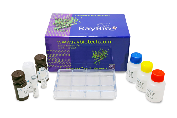
- Human Cytokine Antibody Array C1000 Membranes
- Blocking Buffer
- Wash Buffer 1
- Wash Buffer 2
- Biotinylated Detection Antibody Cocktail
- Streptavidin-Conjugated HRP
- Detection Buffer C
- Detection Buffer D
- Lysis Buffer
- 8-Well Incubation Tray
- Plastic Sheets
- Array Templates
- Manual
- Pipettors, pipet tips and other common lab consumables
- Orbital shaker or oscillating rocker
- Tissue Paper, blotting paper or chromatography paper
- Adhesive tape or Saran Wrap
- Distilled or de-ionized water
- A chemiluminescent blot documentation system (such as UVP's ChemiDoc-It® or EpiChem II Benchtop Darkroom or GE's ImageQuant™ LAS 4000 or Amersham Imagers 600 and 680), X-ray Film and a suitable film processor, or another chemiluminescent detection system.
- Block membranes
- Incubate with Sample
- Incubate with Biotinylated Detection Antibody Cocktail
- Incubate with HRP-Conjugated Streptavidin
- Incubate with Detection Buffers
- Image with chemiluminescent imaging system
- Perform densitometry and analysis
Storage/Stability
Celis JE, Moreira JMA, Cabezón T, Gromov P, et al. Identification of Extracellular and Intracellular Signaling Components of the Mammary Adipose Tissue and Its Interstitial Fluid in High Risk Breast Cancer Patients: Toward Dissecting The Molecular Circuitry of Epithelial-Adipocyte Stromal Cell Interactions. Mol Cell Proteomics. 2005;4:492-522.
Celis JE, Gromov P, Moreira JMA, Cabezon T, et al. Apocrine Cysts of the Breast: Biomarkers, Origin, Enlargement, and Relation with Cancer Phenotype. Mol Cell Proteomics. 2006;5:432-483.
Moro C, Klimeakova E, Lolmede K, Berlan M, et al. Atrial natriuretic peptide inhibits the production of adipokines and cytokines linked to inflammation and insulin resistance in human subcutaneous adipose tissue. Diabetologia. 2007; 50:1038-1047.
Broux B., Pannemans K., Zhang X., et al. CX(3)CR1 drives cytotoxic CD4(+)CD28(-) T cells into the brain of multiple sclerosis patients. J Autoimmun. 2012 Feb;38(1):10-9. doi: 10.1016/j.jaut.2011.11.006
Wiley C., Velarde M., Lecot P., et al. Mitochondrial Dysfunction Induces Senescence with a Distinct Secretory Phenotype. Cell Metab . 2016 Feb 9;23(2):303-14. doi: 10.1016/j.cmet.2015.11.011. Epub 2015 Dec 10.
Henrichot E, Juge-Aubry CE, Pernin A, Pache J-C, et al. Production of Chemokines by Perivascular Adipose Tissue: A Role in the Pathogenesis of Atherosclerosis? Aterioscler Thromb Vasc Biol. 2005;25:2594-2599
Lee J., Schierer S., Blume K., et al. HIV-Nef and ADAM17-Containing Plasma Extracellular Vesicles Induce and Correlate with Immune Pathogenesis in Chronic HIV Infection. EBioMedicine. 2016 Apr; 6: 103-113.
Loria F., Diaz-Nido J. Frataxin knockdown in human astrocytes triggers cell death and the release of factors that cause neuronal toxicity. Neurobiology of Disease, 76 (2015) 1-12. doi:10.1016/j.nbd.2014.12.017
Boucek RJ., et al. < i> Ex Vivo Paracrine Properties of Cardiac Tissue: Effects of Chronic Heart Failure. J of Heart and Lung Transpl. Available online 17 July 2014 DOI: 10.1016/j.healun.2014.07.010
Shi Z., Yang YM., Chen LP., et al. Enhanced chemosensitization in multidrug-resistant human breast cancer cells by inhibition of IL-6 and IL-8 production. Breast Cancer Res Treat. 2012 Oct;135(3):737-47. doi: 10.1007/s10549-012-2196-0
-
reliable and excellent cytokine arrayI used these arrays to identified secreted factors from my cultured conditioned media from co-culture of immune cells with prostate cancer cells. The products gave me very consistent results. In addition to identification of the targets, it also allows quantifying the secreted factors from the conditions that I want to compare.
from Clark Atlanta University,
on
Great productUsed to probe supernatant from cultured microglia. Blots came out with higher background than expected but otherwise easy to use.from University of Georgia,
on
Great productI used this array for the second time to study the effect of my compound of different cytokine expression. I published the paper recently:Messeha Messeha SS, Zarmouh NO, Mendonca P, Carolyn Cotton, Karam F A Soliman: Molecular mechanism of gossypol mediating CCL2 and IL‑8 attenuation in triple‑negative breast cancer cells. Molecular medicine reports 22: 1213-1226, 2020.from Florida A & M University, Tallahassee, Florida,
on
Great productEasy, reliable and replicablefrom Penn State,
on
Great productThis is a very trustworthy product.from University of Southern California,
on
Great productI have been using these arrays and having pretty good results. My different treatments up and down regulate the expression of these cytokines, which can be clearly seen on the membrane spots.from Florida A & M University,
on
Great productI used this product for testing tumor tissues protein lysates to compare cytokine between control and treated conditions. The results so far has been satisfactoryon
Write Your Own ReviewAsk a Question