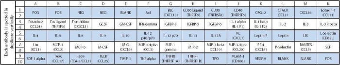
RayBio® C-Series Mouse Cytokine Antibody Array 3 Kit. Detects 62 Mouse Cytokines. Suitable for all liquid sample types.

Product Description
Specifications
| Size | 2 Sample Kit, 4 Sample Kit, 8 Sample Kit |
|---|---|
| Species | Mouse |
| Quantitative/Semi-Quantitative | Semi-Quantitative |
| Number of Targets Detected | 62 |
| Compatible Sample Types | Cell Culture Supernatants, Plasma, Serum, Tissue Lysates, Cell Lysates |
| Solid Support | Membrane |
| Method Of Detection | Chemiluminescence |
| Design Principle | Sandwich-based |
| Estimated Lead Time | 1-2 business days |
| Shipping Type | Blue ice |
| Storage | -20°C |
Risk-Free Guarantee
We offer a 100% guarantee on all ELISA kits and membrane cytokine arrays.
Learn More
Amazon Gift Cards!
$5 Amazon gift card in every kit box purchased.
| Scroll over each target protein for more information | ||||
|---|---|---|---|---|
|
Axl
|
BLC
(CXCL13)
|
CD30 Ligand
(TNFSF8)
|
CD30
(TNFRSF8)
|
CD40
(TNFRSF5)
|
|
CRG-2
|
CTACK
(CCL27)
|
CXCL16
|
Eotaxin-1
(CCL11)
|
Eotaxin-2
(MPIF-2/CCL24)
|
|
Fas Ligand
(TNFSF6)
|
Fractalkine
(CX3CL1)
|
GCSF
|
GM-CSF
|
IFN-gamma
|
|
IGFBP-3
|
IGFBP-5
|
IGFBP-6
|
IL-1 beta
(IL-1 F2)
|
IL-10
|
|
IL-12 p40/p70
|
IL-12 p70
|
IL-13
|
IL-17A
|
IL-1 alpha
(IL-1 F1)
|
|
IL-2
|
IL-3
|
IL-3 R beta
|
IL-4
|
IL-5
|
|
IL-6
|
IL-9
|
KC
(CXCL1)
|
Leptin
|
Leptin R
|
|
LIX
|
L-Selectin
(CD62L)
|
Lymphotactin
(XCL1)
|
MCP-1
(CCL2)
|
MCP-5
|
|
M-CSF
|
MIG
(CXCL9)
|
MIP-1 alpha
(CCL3)
|
MIP-1 gamma
|
MIP-2
|
|
MIP-3 beta
(CCL19)
|
MIP-3 alpha
(CCL20)
|
Platelet Factor 4
(CXCL4)
|
P-Selectin
|
RANTES
(CCL5)
|
|
SCF
|
SDF-1 alpha
(CXCL12 alpha)
|
TNF RI
(TNFRSF1A)
|
TNF RII
(TNFRSF1B)
|
TARC
(CCL17)
|
|
I-309
(TCA-3/CCL1)
|
TECK
(CCL25)
|
TIMP-1
|
TNF alpha
|
Thrombopoietin
(TPO)
|
|
VCAM-1
(CD106)
|
VEGF-A
|
|||
Species Detected
Application Notes
- Mouse Cytokine Antibody Array C3 Membranes
- Blocking Buffer
- Wash Buffer 1
- Wash Buffer 2
- Biotinylated Detection Antibody Cocktail
- Streptavidin-Conjugated HRP
- Detection Buffer C
- Detection Buffer D
- Lysis Buffer
- 8-Well Incubation Tray
- Plastic Sheets
- Array Templates
- Manual
- Pipettors, pipet tips and other common lab consumables
- Orbital shaker or oscillating rocker
- Tissue Paper, blotting paper or chromatography paper
- Adhesive tape or Saran Wrap
- Distilled or de-ionized water
- A chemiluminescent blot documentation system (such as UVP's ChemiDoc-It® or EpiChem II Benchtop Darkroom or GE's ImageQuant™ LAS 4000 or Amersham Imagers 600 and 680), X-ray Film and a suitable film processor, or another chemiluminescent detection system.
- Block membranes
- Incubate with Sample
- Incubate with Biotinylated Detection Antibody Cocktail
- Incubate with HRP-Conjugated Streptavidin
- Incubate with Detection Buffers
- Image with chemiluminescent imaging system
- Perform densitometry and analysis
Storage/Stability
Pelaez-Garcia A., Barderas R., Batlle R., et al. A proteomic analysis reveals that Snail regulates the expression of the nuclear orphan receptor Nuclear Receptor Subfamily 2 Group F Member 6 (Nr2f6) and interleukin 17 (IL-17) to inhibit adipocyte differentiation. Mol Cell Proteomics. 2015 Feb;14(2):303-15. doi: 10.1074/mcp.M114.045328.
Fuster-Matanzo, Almudena, et al. "Dual effects of increased glycogen synthase kinase-3? activity on adult neurogenesis." Human molecular genetics 22.7 (2012): 1300-1315.
Nair, Ashwin, et al. "Biomaterial implants mediate autologous stem cell recruitment in mice." Acta biomaterialia 7.11 (2011): 3887-3895.
Kang TW., et al. Senescence surveillance of pre-malignant hepatocytes limits liver cancer development. Nature. 2011 Nov 9;479(7374):547-51. doi: 10.1038/nature10599.
Moon H., Donahue L., Choi E., et al. Melanocyte Stem Cell Activation and Translocation Initiate Cutaneous Melanoma in Response to UV Exposure. Cell Stem Cell . 2017 Nov 2;21(5):665-678.e6. doi: 10.1016/j.stem.2017.09.001. Epub 2017 Oct 12.
Hughes, David J., et al. "Chemokine binding protein M3 of murine gammaherpesvirus 68 modulates the host response to infection in a natural host." PLoS Pathog 7.3 (2011): e1001321.
Kang TW., et al. Senescence surveillance of pre-malignant hepatocytes limits liver cancer development. Nature. 2011 Nov 9;479(7374):547-51. doi: 10.1038/nature10599.
Vergori L., Lauret E., Gaceb A., et al. PPAR? Regulates Endothelial Progenitor Cell Maturation and Myeloid Lineage Differentiation Through a NADPH Oxidase-Dependent Mechanism in Mice. Stem Cells. 2015 Apr;33(4):1292-303. doi: 10.1002/stem.1924.
Simon M., Ven Meter M., Ablaeva J., et al. LINE1 Derepression in Aged Wild-Type and SIRT6-Deficient Mice Drives Inflammation. Cell Metab . 2019 Apr 2;29(4):871-885.e5. doi: 10.1016/j.cmet.2019.02.014. Epub 2019 Mar 7.
Szabolcsi V., Celio M. De novo Expression of Parvalbumin in Ependymal Cells in Response to Brain Injury Promotes Ependymal Remodeling and Wound Repair. GLIA 2014, epub ahead of print. DOI: 10.1002/glia.22768
-
Great ProductGreat Product! The protocol was straightforward and overall very user friendly. Our lab used this to analyze mouse lung tissue. We have used this numerous times in the past and will continue to do so in the future.
from Feinstein Institutes for Medical Research,
on
Simple to useThe protocol is straightforward and easy to follow. I used it to analyze mouse liver tissue lysate. You might need to use a couple of membranes to standardize your protocol as too low or too much protein loaded might affect the output. I also found that using the lysis buffer contained in the kit worked better than the NP40 i used earlier.from Howard University,
on
Fantastic ProductWe used to assay pancreatic tissue lysate, for pancreatic cancer project. It's a great product. We are ordering more for our other project on breast cancer too. Thanks.from south Dakota state university,
on
Great productWe used the assay to analyze lung tissue extracts at 500ug per membrane. We got a good signal and reproducible results. However, I would recommend using different ECL reagent for detection, the one provided with the kit is not the best.from University of Kansas Medical Center,
on
Great productThe protocol is easy and user friendly. However, the read out proved difficult as the radiance between samples in different conditions can not be discerned clearly even when using advanced western blot readout software. Tried different exposures and did not get adequate results for a manuscript figure.from National Cancer Institute, NIH,
on
Great productWe used the above kit to get the cytokine patterns in the secretome of the infected bone marrow derived macrophages. The kit seems to be consistent. With in vitro stimulated cells, the cytokine levels were found. We used undiluted samples in analysis. The FBS from VWR at 2% was also present in the medium.from University of Chicago,
on
Great productWe used the AAM-CYT-3-8 Kit. With the detailed protocol manual, we could take all the necessary precautions to get consistent signals with the positive controls and we could confirm the same after repeating it for the selected individual cytokines that were deferentially unregulatedfrom W.M. Keck Center for Transgene Research,
on
Great productThis product is fantastic! We use to analyze the connection between irradiation dose and inflammation in mouse plasma in mice with different genetic backgrounds. The protocol is easy to follow and the results are always clear to interpret.from Roswell Park Cancer Institute,
on
Great productI used this array in the serum samples of deer mice and got good pictures. The images can be easily analyzed by Imagelab.from University of South Carolina,
on
Great productProtocol is easy to follow and convenient to use. It is time-saving.from Purdue University,
on
Great productI used this array to analyze some astrocyte culture-conditioned (serum-free) media (about 1 million cells/well). I didn't use the software for analysis, and it took me about a day to analyze the four membranes. I didn't dilute the media and I certainly wasn't maxing out the signal when going with all of the optional overnight incubations.from Northwestern University,
on
Write Your Own ReviewAsk a Question