
Product Description
Specifications
| Size | 2 Sample Kit, 4 Sample Kit, 8 Sample Kit |
|---|---|
| Species | Human |
| Quantitative/Semi-Quantitative | Semi-Quantitative |
| Number of Targets Detected | 55 |
| Compatible Sample Types | Cell Culture Supernatants, Plasma, Serum, Tissue Lysates, Cell Lysates |
| Solid Support | Membrane |
| Method Of Detection | Chemiluminescence |
| Design Principle | Sandwich-based |
| Research Area | Post-Translational Modifications, Phosphorylation, Akt Signaling, JAK/STAT Signaling, MAPK Signaling, TGF-beta Signaling, NFkB Signaling |
| Estimated Lead Time | 1-2 business days |
| Shipping Type | Blue ice |
| Storage | -20°C |
Risk-Free Guarantee
We offer a 100% guarantee on all ELISA kits and membrane cytokine arrays.
Learn More
Amazon Gift Cards!
$5 Amazon gift card in every kit box purchased.
Related Products

| Scroll over each target protein for more information | ||||
|---|---|---|---|---|
|
4E-BP1 (P-Thr36)
|
AKT (P-Ser473)
|
ATF2 (P-Thr69/71)
|
ATM (P-Ser1981)
|
|
|
BAD (P-Ser112)
|
C-Fos (P-Thr232)
|
CREB (P-Ser133)
|
EGFR (P-Ser1070)
|
|
|
ERK1/2 (P-Thr202/P-Tyr204/P-Tyr185/P-Tyr187)
|
GSK-3 alpha (P-Ser21)
|
GSK-3 beta (P-Ser9)
|
HDAC2 (P-Ser394)
|
|
|
HDAC4 (P-Ser632)
|
HSP27 (P-Ser82)
|
IKBa (P-Ser32)
|
JAK1 (P-Tyr1022)
|
JAK2 (P-Tyr1007/1008)
|
|
JNK (P-Thr183)
|
MEK (P-Ser217/221)
|
MKK3 (P-Ser189)
|
MKK6 (P-Ser207)
(MEK6)
|
MSK1 (P-Ser376)
|
|
MSK2 (P-Ser360)
|
mTOR (P-Ser2448)
|
NF-kB (P-Ser536)
|
p27 (P-Thr198)
|
p38 (P-Thr180/Tyr182)
|
|
P53 (P-Ser15)
|
P70S6K (P-Thr421/Ser424)
|
PDK1 (P-Ser241)
|
PRAS40 (P-Thr246)
|
PTEN (P-Ser380)
|
|
Raf-1 (P-Ser301)
|
RPS6 (P-Ser235/236)
|
RSK1 (P-Ser380)
|
RSK2 (P-Ser386)
|
SHP-1 (P-Ser591)
|
|
SHP2 (P-Tyr542)
|
SMAD1 (P-Ser463/465)
|
SMAD2 (P-Ser245/250/255)
|
SMAD4 (P-Thr277)
|
SMAD5 (P-Ser463/465)
|
|
Src (P-Tyr419)
|
STAT1 (P-Ser727)
|
STAT2 (P-Tyr689)
|
STAT3 (P-Tyr705)
|
STAT5 (P-Tyr694)
|
|
STAT6 (P-Tyr641)
|
TAK1 (P-Ser412)
|
TBK1 (P-Ser172)
|
TYK2 (P-Tyr1054)
|
ZAP70 (P-Tyr292)
|
Application Notes
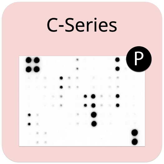
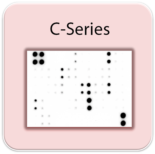
- Human Phosphorylation Pathway Profiling Array C55 Membranes
- Blocking Buffer
- Detection Antibody Cocktail
- 500X HRP-Anti-Rabbit IgG Concentrate
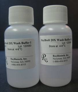
- 20X Wash Buffer I Concentrate
- 20X Wash Buffer II Concentrate
- 2X Cell Lysis Buffer Concentrate
- Detection Buffer C
- Detection Buffer D
- 8-Well Incubation Tray w/ Lid
- Protease Inhibitor Cocktail
- 100x Phosphatase Inhibitor Cocktail I
- Phosphatase Inhibitor Cocktail II
- Plastic Sheets
- Array Map Template
- Manual
- Pipettors, pipet tips and other common lab consumables
- Orbital shaker or oscillating rocker
- Tissue Paper, blotting paper or chromatography paper
- Adhesive tape or Saran Wrap
- Distilled or de-ionized water
- A chemiluminescent blot documentation system (such as UVP's ChemiDoc-It® or EpiChem II Benchtop Darkroom or GE's ImageQuant™ LAS 4000 or Amersham Imagers 600 and 680), X-ray Film and a suitable film processor, or another chemiluminescent detection system.
- Block membranes
- Incubate with Sample
- Incubate with Detection Antibody Cocktail
- Incubate with HRP-Conjugated anti-IgG
- Incubate with Detection Buffers
- Image with chemiluminescent imaging system
- Perform densitometry and analysis
Storage/Stability
Zhang, Dan, et al. "MALAT1 is involved in the pathophysiological process of PCOS by modulating TGF? signaling in granulosa cells." Molecular and cellular endocrinology 499 (2020): 110589.
Zhang D, Tang HY, Tan L, Zhao DM. MALAT1 is involved in the pathophysiological process of PCOS by modulating TGF? signaling in granulosa cells. Mol Cell Endocrinol. 2020 Jan 1;499:110589. doi: 10.1016/j.mce.2019.110589. Epub 2019 Sep 23. PMID: 31557499.
-
Great system, easy to use, would use againThis kit worked really well. The kit came with everything you would need to run the experiment. I used the minimum recommended amount of protein and it worked very well. I did have a couple issues, but that wouldn't stop me from ordering it again. First, I tried marking the membranes at the their recommendation, but the ink I used bled onto a few of the protein dots so just be aware that it may happen. Also, the analysis software was a bit of pain. For each pathway, there was a specific excel file (so 5 total) and some were updated and some were not. It would have been easier to have everything in one file that also didn't have several tabs for the analysis, or at least a much more organized file with clear description of each step. I ended up doing the analysis on my own after struggling for a bit. But overall, very happy with my purchase.
on
Irwineasy to use, with clear results at the end. I was using human fibroblasts to investigate the affect of Crohn's disease serum on downstream pathways, and worked well with less than recommended protein concentration due to low concentration from the sampleson
Great productIt was easy to test multiple phosphorylation targets with limited sample quantity and workflow was really easy and uncomplicated. I used primary human endothelium cells with 100 ug of protein for each array and signals were good with 10-sec exposure.from University of Wisconsin-Madison,
on
Great productThis kit works pretty well for my samples. I used 700 ug of my samples (cell lysates) and performed exactly according to the instructional protocol and I got a very strong signal. The only reason that I didn't give it 5 stars was we had to purchase their software to analyze the results. As we already spent nearly $1000, we should be able to use the software for free, at least one time free.from Kalya Pharma,
on
Write Your Own Review