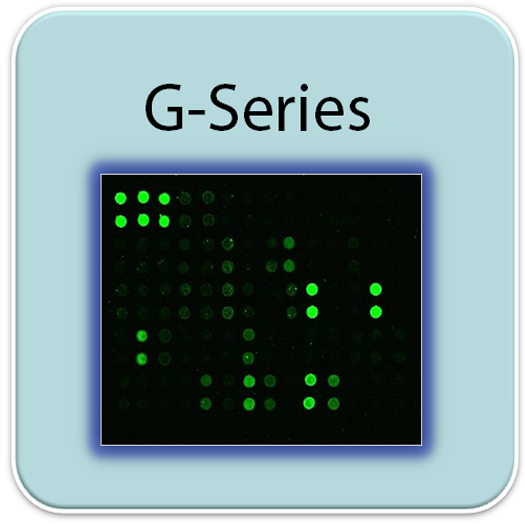
RayBio® G-Series Human Apoptosis Antibody Array 1 Kit. Detects 43 Human Apoptotic Factors. Suitable for all liquid sample types but intended for use with cell and tissue lysates.
Product Description
Specifications
| Size | 4 Sample Kit, 8 Sample Kit |
|---|---|
| Species | Human |
| Quantitative/Semi-Quantitative | Semi-Quantitative |
| Number of Targets Detected | 43 |
| Compatible Sample Types | Cell Culture Supernatants, Plasma, Serum, Tissue Lysates, Cell Lysates |
| Solid Support | Glass Slide |
| Method Of Detection | Fluorescence Laser Scanner |
| Design Principle | Sandwich-based |
| Research Area | Apoptosis |
| Estimated Lead Time | 1-2 business days |
| Shipping Type | Blue ice |
| Storage | -20°C |

Amazon Gift Cards!
$5 Amazon gift card in every kit box purchased.
| Scroll over each target protein for more information | ||||
|---|---|---|---|---|
bad
| bax
| bcl-2
| bcl-w
| BID
|
BIM
| Caspase-3
| Caspase-8
| CD40 (TNFRSF5)
| CD40 Ligand (TNFSF5)
|
cIAP-2
| Cytochrome C | DR6 (TNFRSF21)
| Fas (TNFRSF6/Apo-1)
| Fas Ligand (TNFSF6)
|
HSP27
| HSP60
| HSP70
| HTRA2
| IGF-1
|
IGF-2
| IGFBP-1
| IGFBP-2
| IGFBP-3
| IGFBP-4
|
IGFBP-5
| IGFBP-6
| IGF-1 R
| livin
| p21
|
p27
| p53
| SMAC
| Survivin (BIRC5)
| TNF RI (TNFRSF1A)
|
TNF RII (TNFRSF1B)
| TNF alpha
| TNF beta (TNFSF1B)
| TRAIL R1 (TNFRSF10A/DR4)
| TRAIL R2 (TNFRSF10B/DR5)
|
TRAIL R3 (TNFRSF10C)
| TRAIL R4 (TNFRSF10D)
| XIAP
| ||
Application Notes
- Human Apoptosis Antibody Array G1 Slide(s)
- Blocking Buffer
- Wash Buffer 1
- Wash Buffer 2
- Biotinylated Detection Antibody Cocktail
- Streptavidin-Conjugated Fluor
- Lysis Buffer
- Protease Inhibitor Cocktail
- Adhesive Plastic Strips
- 30 ml Centrifuge Tube
- Manual
- Small plastic boxes or containers
- Pipettors, pipette tips and other common lab consumables
- Orbital shaker or oscillating rocker
- Aluminum foil
- Gene microarray scanner or similar laser fluorescence scanner
View Compatible Laser Scanners
Don't have a compatible scanner? RayBiotech now offers FREE scanning service for all RayBio glass slide antibody arrays! Learn More
- Dry the glass slide
- Block array surface
- Incubate with Sample
- Incubate with Biotinylated Detection Antibody Cocktail
- Incubate with Streptavidin-Conjugated Fluor
- Disassemble the glass slide
- Scan with a gene microarray laser scanner
- Perform densitometry and analysis
Storage/Stability
The Presence of HIV-1 Tat Second Exon Delays Fas-Mediated Apoptosis in CD4+ T lymphocytes: a Potential Mechanism for Persistent Viral Production
López-Huertas, MarÃa Rosa, et al. "The Presence of HIV-1 Tat Protein Second Exon Delays Fas Protein-mediated Apoptosis in CD4+ T Lymphocytes A POTENTIAL MECHANISM FOR PERSISTENT VIRAL PRODUCTION." Journal of Biological Chemistry 288.11 (2013): 7626-7644.
Qin M., Luo Y., Meng X., et al. Myricitrin attenuates endothelial cell apoptosis to prevent atherosclerosis: An insight into PI3K/Akt activation and STAT3 signaling pathways. Vascul Pharmacol. 2015 Jul;70:23-34. doi: 10.1016/j.vph.2015.03.002
Asif M., Shafaei A., Jafari S., et al. Isoledene from Mesua ferrea oleo-gum resin induces apoptosis in HCT116 cells through ROS-mediated modulation of multiple proteins in the apoptotic pathways: A mechanistic study. Toxicol Lett. 2016 Aug 22;257:84-96. doi: 10.1016/j.toxlet.2016.05.027.
Ibrahim M., et al. ?-Mangostin from Cratoxylum arborescens demonstrates apoptogenesis in MCF-7 with regulation of NF-?B and Hsp70 protein modulation in vitro, and tumor reduction in vivo. Drug Des Devel Ther. 2014 Sep 27;8:1629-47. doi: 10.2147/DDDT.S66105
Li X., Liu X., Xu Y., et al. Expression profile of apoptotic and proliferative proteins in hypoxic HUVEC treated with statins. Int J Oncol. 2015 Feb;46(2):677-84. doi: 10.3892/ijo.2014.2780.
Chen, Yao, et al. "WAP four-disulfide core domain protein 2 gene (WFDC2) is a target of estrogen in ovarian cancer cells." Journal of ovarian research 9.1 (2016): 10.
Sidhu P., et al. Anticancer activity of VDR-coregulator inhibitor PS121912. Cancer Chemotherapy and Pharmacology Published Online August 2014. DOI 10.1007/s00280-014-2549-y
Mohidin T., Ng C. BARF1 gene silencing triggers caspase-dependent mitochondrial apoptosis in Epstein-Barr virus-positive malignant cells. J Biosci. 2015 Mar;40(1):41-51.
Qin Y., Zhang S., Den S., et al. Epigenetic silencing of miR-137 induces drug resistance and chromosomal instability by targeting AURKA in multiple myeloma. Leukemia. 2017 Jan 6. doi: 10.1038/leu.2016.325.
-
Great productMonitored protein expression in HMCB cells. Company’s free scanning of the array allowed us to use this tool and provide quality research experiences for undergraduates.
from Hiram College,
on
Great productA great assay for a fairly wide array of apoptosis proteins and if you lack the proper scanners the company will quickly scan and analyze your samples at no cost.from Columbia University,
on
Great productA very high-throughput, sensitive product to detect the apoptosis.on
Write Your Own Review
