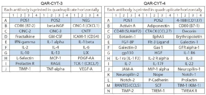
Quantibody® Rat Cytokine Array 67 Kit: A combination of 2 non-overlapping arrays (QAR-CYT-3 (27) & QAR-CYT-4 (40)) to quantitatively measure the concentration of 67 rat cytokines. Suitable for all liquid sample types.
Product Description
Specifications
| Size | 8 Sample Kit, 22 Sample Kit, 50 Sample Kit |
|---|---|
| Species | Rat |
| Quantitative/Semi-Quantitative | Quantitative |
| Number of Targets Detected | 67 |
| Compatible Sample Types | Cell Culture Supernatants, Plasma, Serum, Tissue Lysates, Other Body Fluids, Cell Lysates |
| Solid Support | Glass Slide |
| Method Of Detection | Fluorescence Laser Scanner |
| Design Principle | Sandwich-based |
| Research Area | Antibody Drug Development, Biomarker Detection |
| Estimated Lead Time | 1-2 business days |
| Shipping Type | Blue ice |
| Storage | -20°C |

Amazon Gift Cards!
$5 Amazon gift card in every kit box purchased.
| Scroll over each target protein for more information | ||||
|---|---|---|---|---|
|
4-1BB
(TNFRSF9/CD137)
|
Activin A
|
Adiponectin
(ACRP30)
|
beta-NGF
|
CD48
(SLAMF2)
|
|
CD80
(B7-1)
|
CD86
(B7-2)
|
CINC-1
(CXCL1)
|
CINC-2
|
CINC-3
|
|
CNTF
|
CTACK
(CCL27)
|
Decorin
|
Eotaxin-1
(CCL11)
|
EphA5
|
|
Erythropoietin
|
FGF-BP
|
Flt-3 Ligand
|
Fractalkine
(CX3CL1)
|
Galectin-1
|
|
Galectin-3
|
Gas 1
|
GFR alpha-1
(GDNF R)
|
GM-CSF
|
gp130
|
|
HGF
|
ICAM-1
(CD54)
|
IFN-gamma
|
IL-1 alpha
(IL-1 F1)
|
IL-1 beta
(IL-1 F2)
|
|
IL-1 R6
(IL-1 Rrp2)
|
IL-1 Ra
(IL-1 F3)
|
IL-2
|
IL-2 R alpha
|
IL-3
|
|
IL-4
|
IL-6
|
IL-7
|
IL-10
|
IL-13
|
|
IL-17F
|
IL-22
|
JAM-A
(CD321/F11R)
|
LIX
|
L-Selectin
(CD62L)
|
|
MCP-1
(CCL2)
|
MIP-1 alpha
(CCL3)
|
Neuropilin-1
|
Neuropilin-2
|
Nope
|
|
Notch-1
|
Notch-2
|
P-Cadherin
|
PDGF-AA
|
Prolactin
|
|
Prolactin R
|
RAGE
|
RANTES
(CCL5)
|
SCF
|
TCK-1
(CXCL7)
|
|
TIM-1
(KIM-1)
|
TIMP-1
|
TIMP-2
|
TNF alpha
|
TREM-1
|
|
TWEAK R
(TNFRSF12)
|
VEGF-A
|
|||
Species Detected
Rat
Application Notes
Suggested Application
Multiplexed Protein Detection; Biomarker Screening; Identifying Key Factors; Confirming Biological Process; Biomarker Validation; Validation of Antibody Array Results; Quantitative Protein Detection; Establishing Normal Range
Kit Components
- Rat Cytokine Array Q3 Slide(s); Rat Cytokine Array Q4 Slide(s)
- Blocking Buffer
- Wash Buffer 1
- Wash Buffer 2
- Lyophilized Standard Mix
- Biotinylated Detection Antibody Cocktails
- Streptavidin-Conjugated Fluor
- Slide Washer/Dryer
- Adhesive Plastic Strips
- Manual
Other Materials Required
- Distilled or deionized water
- Small plastic boxes or containers
- Pipettors, pipette tips and other common lab consumables
- Orbital shaker or oscillating rocker
- Aluminum foil
- Gene microarray scanner or similar laser fluorescence scanner
View Compatible Laser Scanners
Don't have a compatible scanner? RayBiotech now offers FREE scanning service for all RayBio glass slide antibody arrays! Learn More
Protocol Outline
- Dry the glass slide
- Prepare Standards
- Block array surface
- Incubate with Samples and Standards
- Incubate with Biotinylated Detection Antibody Cocktail
- Incubate with Streptavidin-Conjugated Fluor
- Disassemble the glass slide
- Scan with a gene microarray laser scanner
- Perform densitometry and analysis
Storage/Stability
For best results, store the entire kit frozen at -20°C upon arrival. Stored frozen, the kit will be stable for at least 6 months which is the duration of the product warranty period. Once thawed, store array slide(s), standard mix, detection antibody cocktail, and Cy3-Conjugated Streptavidin at -20°C and all other reagents undiluted at 4°C for no more than 3 months.
Luck C., Demarco V., Mahmood A., et al. Differential Regulation of Cardiac Function and Intracardiac Cytokines by Rapamycin in Healthy and Diabetic Rats. Oxid Med Cell Longev. 2017;2017:5724046. doi: 10.1155/2017/5724046
Species:
Rat
Sample Type:
Tissue Lysate (Heart Tissue lysate from diabetic rats)
Wan, L., Wang, Y., Tang, Y. et al. Gefitinib-Induced Cutaneous Toxicities in Brown Norway Rats Are Associated with Macrophage Infiltration. Inflammation 43, 2137-2146 (2020). https://doi.org/10.1007/s10753-020-01281-3
Species:
Rat
Sample Type:
Tissue Lysate (skin)
Cheng XQ, Liang XZ, Wei S, Ding X, Han GH, Liu P, Sun X, Quan Q, Tang H, Zhao Q, Shang AJ, Peng J (2020) Protein microarray analysis of cytokine expression changes in distal stumps after sciatic nerve transection. Neural Regen Res 15(3):503-511. doi:10.4103/1673-5374.266062
Species:
Rat
Sample Type:
Tissue Lysate (extracted from distal nerve stumps of sciatic nerves)
Fischer G., Wang F., Xiang H., et al. Inhibition of neuropathic hyperalgesia by intrathecal bone marrow stromal cells is associated with alteration of multiple soluble factors in cerebrospinal fluid. Exp Brain Res. 2017 Sep;235(9):2627-2638. doi: 10.1007/s00221-017-5000-x.
Species:
Rat
Sample Type:
CSF (CSF relief of pain when injected)
Write Your Own Review