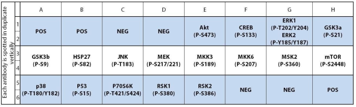
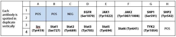
RayBio® Mouse JAK/STAT Pathway Phosphorylation Array is a multiplex assay on nitrocellulose membrane for the simultaneous identification of 12 phosphorylated mouse proteins in cell and tissue lysates.
The JAK/STAT pathway is one of the most important signaling pathways that link extracellular signals to the machinery that controls fundamental cellular processes such as immunity, proliferation, differentiation, apoptosis and oncogenesis.
The RayBio® Mouse JAK/STAT Pathway Phosphorylation Array Kit is a rapid, sensitive, and economical tool for the simultaneous detection of the relative levels of phosphorylation of 12 JAK/STAT pathway proteins. Each antibody was carefully validated using appropriate cell lysates. Using the RayBio® Mouse JAK/STAT Pathway Phosphorylation Array Kit, you can verify multiple specific targets activation in your system without spending excess time and effort in performing an analysis of ELISA and/or Western Blot. The RayBio® Mouse JAK/STAT Pathway Phosphorylation Array Kit has several advantages over the detection of JAK/STAT pathway markers using single-target ELISA kits or Western blots:
More Data, Same or Less Sample: Antibody arrays provide high-content screening using about the same sample volume as traditional ELISA.
Global View of Protein Expression: Antibody array screening improves the chances for discovering key factors, disease mechanisms, or biomarkers related to cell signaling.
Similar (sometimes better) Sensitivity: As little as 4 pg/ml of MCP-1 can be detected using the C-Series array format. In contrast, our similar MCP-1 ELISA assay has a sensitivity of 40 pg/ml of MCP-1.
Wider Detection Range: ELISA typically detects a concentration range of 100 to 1000-fold, while RayBiotech arrays can detect concentration ranges up to 10,000-fold.
Better Precision: As determined by densitometry, the inter-array Coefficient of Variation (CV) of spot signal intensities is 5-10%, comparing favorably with ELISA testing (CV = 10-15%).
Product Features
Easy to use
No specialized equipment needed
Compatible with nearly any liquid sample
Proven technology (many publications)
Highly sensitive (pg/ml)
Sandwich ELISA specificity
Higher density than ELISA, Western blot or bead-based multiplex
Multiplexed Protein Detection; Detection of Relative Protein Expression; Detecting Patterns of Cytokine Expression; Biomarker/ Key Factor Screening; Identifying Key Factors; Confirming a Biological Process
Pipettors, pipet tips and other common lab consumables
Orbital shaker or oscillating rocker
Tissue Paper, blotting paper or chromatography paper
Adhesive tape or Saran Wrap
Distilled or de-ionized water
A chemiluminescent blot documentation system (such as UVP's ChemiDoc-It® or EpiChem II Benchtop Darkroom or GE's ImageQuant™ LAS 4000 or Amersham Imagers 600 and 680), X-ray Film and a suitable film processor, or another chemiluminescent detection system.
Protocol Outline
Block membranes
Incubate with Sample
Incubate with Detection Antibody Cocktail
Incubate with HRP-Conjugated anti-IgG
Incubate with Detection Buffers
Image with chemiluminescent imaging system
Perform densitometry and analysis
Storage/Stability
For best results, store the entire kit frozen at -20°C upon arrival. Stored frozen, the kit will be stable for at least 6 months which is the duration of the product warranty period. Once thawed, store array membranes at -20°C and all other reagents undiluted at 4°C for no more than 3 months.