Human JAK/STAT Pathway Phosphorylation Array C1
C-Series Human JAK/STAT Pathway Phosphorylation Array 1 Kit. Detects 12 phosphorylated or cleaved human Apoptotic Factors. Suitable for all liquid sample types but intended for use with cell and tissue lysates.
Product Description
Specifications
| Size | 2 Sample Kit, 4 Sample Kit, 8 Sample Kit |
|---|---|
| Species | Human |
| Quantitative/Semi-Quantitative | Semi-Quantitative |
| Number of Targets Detected | 12 |
| Compatible Sample Types | Cell Culture Supernatants, Plasma, Serum, Tissue Lysates, Cell Lysates |
| Solid Support | Membrane |
| Method Of Detection | Chemiluminescence |
| Design Principle | Sandwich-based |
| Research Area | Post-Translational Modifications, Phosphorylation, Akt Signaling, HER/ErbB Signaling, JAK/STAT Signaling, MAPK Signaling, PI3K-AKT Signaling, Notch Signaling |
| Estimated Lead Time | 1-2 business days |
| Shipping Type | Blue ice |
| Storage | -20°C |
Risk-Free Guarantee
We offer a 100% guarantee on all ELISA kits and membrane cytokine arrays.
Learn More
Amazon Gift Cards!
$5 Amazon gift card in every kit box purchased.
Related Products

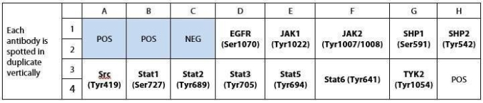
| Scroll over each target protein for more information | ||||
|---|---|---|---|---|
|
EGFR (P-Ser1070)
|
JAK1 (P-Tyr1022)
|
JAK2 (P-Tyr1007/1008)
|
SHP-1 (P-Ser591)
|
SHP2 (P-Tyr542)
|
|
Src (P-Tyr419)
|
STAT1 (P-Ser727)
|
STAT2 (P-Tyr689)
|
STAT3 (P-Tyr705)
|
STAT5 (P-Tyr694)
|
|
STAT6 (P-Tyr641)
|
TYK2 (P-Tyr1054)
|
|||
Typical Results

Figure 1: A431 cells were grown to 80% confluency and then serum starved overnight. Cells were either untreated (bottom panel) or treated (top panel) with EGF for 20 minutes. Data shown are from a 20 second exposure using a chemiluminescence imaging system.
Application Notes
Suggested Application
Multiplexed Protein Detection; Detection of Relative Protein Expression; Detecting Patterns of Cytokine Expression; Biomarker/ Key Factor Screening; Identifying Key Factors; Confirming a Biological Process
Kit Components
- Human JAK/STAT Pathway Phosporylation Array C1 Membranes
- Blocking Buffer
- Detection Antibody Cocktail
- 1,000X HRP-Anti-Rabbit IgG Concentrate
- 20X Wash Buffer I Concentrate
- 20X Wash Buffer II Concentrate
- 2X Cell Lysis Buffer Concentrate
- Detection Buffer C
- Detection Buffer D
- 8-Well Incubation Tray w/ Lid
- Protease Inhibitor Cocktail
- 100x Phosphatase Inhibitor Cocktail I
- Phosphatase Inhibitor Cocktail II
- Plastic Sheets
- Array Map Template
- Manual
Other Materials Required
- Pipettors, pipet tips and other common lab consumables
- Orbital shaker or oscillating rocker
- Tissue Paper, blotting paper or chromatography paper
- Adhesive tape or Saran Wrap
- Distilled or de-ionized water
- A chemiluminescent blot documentation system (such as UVP's ChemiDoc-It® or EpiChem II Benchtop Darkroom or GE's ImageQuant™ LAS 4000 or Amersham Imagers 600 and 680), X-ray Film and a suitable film processor, or another chemiluminescent detection system.
Protocol Outline
- Block membranes
- Incubate with Sample
- Incubate with Detection Antibody Cocktail
- Incubate with HRP-Conjugated anti-IgG
- Incubate with Detection Buffers
- Image with chemiluminescent imaging system
- Perform densitometry and analysis
Storage/Stability
For best results, store the entire kit frozen at -20°C upon arrival. Stored frozen, the kit will be stable for at least 6 months which is the duration of the product warranty period. Once thawed, store array membranes at -20°C and all other reagents undiluted at 4°C for no more than 3 months.
Zheng, Shutao, et al. "NME4 modulates PD-L1 expression via the STAT3 signaling pathway in squamous cell carcinoma." Biochemical and Biophysical Research Communications (2020).
Species:
Human
Sample Type:
Cell Lysate (Squamous cell carcinoma)
-
Really good assayIt worked nicely in human proximal tubule cells. Really easy to use. Great alternative for WB, data can be generated in one day. Very useful for preliminary data for grant application.
from UAB,
on
Write Your Own Review