
Product Description
Specifications
| Size | 2 Sample Kit, 4 Sample Kit, 8 Sample Kit |
|---|---|
| Species | Human, Mouse |
| Quantitative/Semi-Quantitative | Semi-Quantitative |
| Number of Targets Detected | 17 |
| Compatible Sample Types | Cell Culture Supernatants, Plasma, Serum, Tissue Lysates, Cell Lysates |
| Solid Support | Membrane |
| Method Of Detection | Chemiluminescence |
| Design Principle | Sandwich-based |
| Research Area | Post-Translational Modifications, Phosphorylation, Akt Signaling, HER/ErbB Signaling, IGF Signaling, JAK/STAT Signaling, MAPK Signaling, PI3K-AKT Signaling, Wnt/beta-Catenin Signaling, NFkB Signaling |
| Estimated Lead Time | 1-2 business days |
| Shipping Type | Blue ice |
| Storage | -20°C |
Risk-Free Guarantee
We offer a 100% guarantee on all ELISA kits and membrane cytokine arrays.
Learn More
Amazon Gift Cards!
$5 Amazon gift card in every kit box purchased.
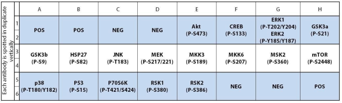
| Scroll over each target protein for more information | ||||
|---|---|---|---|---|
|
Akt (P-Ser473)
(PKB (P-Ser473))
|
CREB (P-Ser133)
|
ERK1 (P-T202/Y204)/ERK2 (P-Y185/Y187)
|
GSK3a (P-Ser21)
|
GSK3b (P-Ser9)
|
|
HSP27 (P-Ser82)
|
JNK (P-Thr183)
|
MEK (P-Ser217/Ser221)
|
MKK3 (P-Ser189)
|
MKK6 (P-Ser207)
|
|
MSK2 (P-Ser360)
|
mTOR (P-Ser2448)
|
p38 (P-Thr180/Tyr182)
|
P53 (P-Ser15)
|
P70S6K (P-Thr421/Ser424)
|
|
RSK1 (P-Ser380)
|
RSK2 (P-Ser386)
|
|||
Application Notes
- Human/Mouse MAPK Phosphorylation Antibody Array C1 Membranes
- Blocking Buffer
- Detection Antibody Cocktail
- 1,000X HRP-Anti-Rabbit-IgG Concentrate
- 20X Wash Buffer I Concentrate
- 20X Wash Buffer II Concentrate
- 2X Cell Lysis Buffer Concentrate
- Detection Buffer C
- Detection Buffer D
- 8-Well Incubation Tray w/ Lid
- Protease Inhibitor Cocktail
- 100x Phosphatase Inhibitor Cocktail I
- Phosphatase Inhibitor Cocktail II
- Plastic Sheets
- Array Map Template
- Manual
- Pipettors, pipet tips and other common lab consumables
- Orbital shaker or oscillating rocker
- Tissue Paper, blotting paper or chromatography paper
- Adhesive tape or Saran Wrap
- Distilled or de-ionized water
- A chemiluminescent blot documentation system (such as UVP??s ChemiDoc-It® or EpiChem II Benchtop Darkroom or GE's ImageQuant™ LAS 4000 or Amersham Imagers 600 and 680), X-ray Film and a suitable film processor, or another chemiluminescent detection system.
- Block membranes
- Incubate with Sample
- Incubate with Detection Antibody Cocktail
- Incubate with HRP-Conjugated anti-IgG
- Incubate with Detection Buffers
- Image with chemiluminescent imaging system
- Perform densitometry and analysis
Typical Data


Storage/Stability
Yu S., Zhong Q., Chen R., et al. CRLF1 promotes malignant phenotypes of papillary thyroid carcinoma by activating the MAPK/ERK and PI3K/AKT pathways. Cell Death Dis. 2018 Mar 7;9(3):371. doi: 10.1038/s41419-018-0352-0.
Yu, Shi-Tong, et al. "CRLF1 promotes malignant phenotypes of papillary thyroid carcinoma by activating the MAPK/ERK and PI3K/AKT pathways." Cell death & disease 9.3 (2018): 1-13.
Poling, Holly M., et al. "Mechanically induced development and maturation of human intestinal organoids in vivo." Nature biomedical engineering 2.6 (2018): 429-442.
Schuett J., Schuett H., Oberoi R., et al. NADPH oxidase NOX2 mediates TLR2/6-dependent release of GM-CSF from endothelial cells. FASEB J. 2017 Mar 8. pii: fj.201600729R. doi: 10.1096/fj.201600729R
Schuett, Jutta, et al. "NADPH oxidase NOX2 mediates TLR2/6?dependent release of GM?CSF from endothelial cells." The FASEB Journal 31.6 (2017): 2612-2624.
Yang Z, Zou W, Pan Y, Yong KT, Li L, Wang X, Liu D, Chen T, Xue D, Lin G. PEGylated CuInS2/ZnS quantum dots inhibit neurite outgrowth by downregulating the NGF/p75NTR/MAPK pathway. Ecotoxicol Environ Saf. 2021 Jan 1;207:111378. doi: 10.1016/j.ecoenv.2020.111378. Epub 2020 Oct 3. PMID: 33022524.
Dhani, Shanel, et al. "Fusaric acid alters Akt and ampk signalling in c57bl/6 mice brain tissue." Food and Chemical Toxicology (2020): 111252.
Sarsour, Ehab H., et al. "Arachidonate 12-lipoxygenase and 12-hydroxyeicosatetraenoic acid contribute to stromal aging-induced progression of pancreatic cancer." Journal of Biological Chemistry 295.20 (2020): 6946-6957.
L'Huillier C, Jarbeau M, Achamrah N, et al. Glutamine, but not Branched-Chain Amino Acids, Restores Intestinal Barrier Function during Activity-Based Anorexia. Nutrients. 2019;11(6):1348. Published 2019 Jun 15. doi:10.3390/nu11061348
Hou J, Cui C, Kim S, Sung C, Choi C. Ginsenoside F1 suppresses astrocytic senescence-associated secretory phenotype. Chem Biol Interact. 2018;283:75-83. doi:10.1016/j.cbi.2018.02.002
-
Great productThis product was great. It was easy to use and provided great results.
from UAMS,
on
Great productThis product is very easy and straightforward to use. I have been using this for a while now.from New York Medical College,
on
Great productShowed robust results in lung cancer cell lines. Planning to order a couple more to use on other cell lines.from JHU,
on
Great productVery easy-to-use, straightforward assay. Great for lung cancer samples.from Dana Farber Cancer Institute,
on
Great productWe compared the MAPK expression patterns in untreated and doxorubicin-treated mouse hearts. Heart muscle tissue extract were assayed at mid-range protein amounts loaded.from George Washington University,
on
Great product1.Experimental conditions and sample typea. In vitro: Human breast cancer cell line (hBCC)b. In vivo: hBCC grew up in nude mouse bone + mouse bone (human/mouse protein mixed)Pros: pAKT1, pCREB, pGSK3b,p70S6K, pRSK1,2, pMEK1, pp38, and pMKK3,6 are good. The experimental process is very easy. Easy to reproduce the resultsCons: pERK1/2 is a signal relatively weak compared to the Cell signaling antibody.from Yale University,
on
Great productThis assay is very straightforward and provides good results for brain samples.from Emory,
on
Great productThese arrays are what I was looking for. They are easy to use, very clean membranes with super sharp results. These arrays are a great second step for large scale analysis such RNAseq.from The University of Iowa,
on
Great productEasy to use and convenient way to look at multiple targets.from Midwestern University,
on
Write Your Own ReviewAsk a Question