Mouse Apoptosis Signaling Pathway Array C1
C-Series Mouse Apoptosis Antibody Array 1 Kit. Detects 17 phosphorylated or cleaved mouse Apoptotic Factors. Suitable for all liquid sample types but intended for use with cell and tissue lysates.
Product Description
Specifications
| Size | 2 Sample Kit, 4 Sample Kit, 8 Sample Kit |
|---|---|
| Species | Mouse |
| Quantitative/Semi-Quantitative | Semi-Quantitative |
| Number of Targets Detected | 17 |
| Compatible Sample Types | Cell Culture Supernatants, Plasma, Serum, Tissue Lysates, Cell Lysates |
| Solid Support | Membrane |
| Method Of Detection | Chemiluminescence |
| Design Principle | Sandwich-based |
| Research Area | Apoptosis, Akt Signaling, HER/ErbB Signaling, HIF-1 alpha Signaling, IGF Signaling, JAK/STAT Signaling, MAPK Signaling, mTOR Signaling, PI3K-AKT Signaling, TGF-beta Signaling, p53 Signaling, NFkB Signaling, PKC Signaling |
| Estimated Lead Time | 1-2 business days |
| Shipping Type | Blue ice |
| Storage | -20°C |
Risk-Free Guarantee
We offer a 100% guarantee on all ELISA kits and membrane cytokine arrays.
Learn More
Amazon Gift Cards!
$5 Amazon gift card in every kit box purchased.
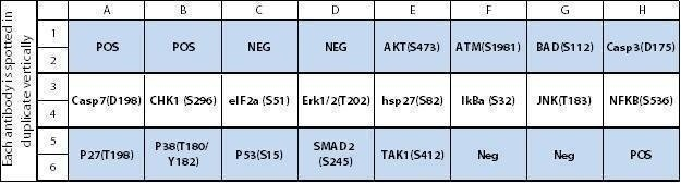
| Scroll over each target protein for more information | ||||
|---|---|---|---|---|
|
AKT (P-Ser473)
|
ATM (P-Ser1981)
|
BAD (P-Ser112)
|
Caspase-3 (P-Asp175)
|
Caspase-7 (P-Asp198)
|
|
CHK1 (P-Ser296)
|
eIF-2a (P-Ser52)
|
ERK1/2 (P-Thr202)
|
HSP27 (P-Ser82)
|
IKBa (P-Ser32)
|
|
JNK (P-Thr183)
|
NF-kB P65 (P-Ser536)
|
p27 (P-Thr198)
|
p38 (P-Thr180/Tyr182)
|
P53 (P-Ser15)
|
|
SMAD2 (P-Ser245)
|
TAK1 (P-Ser412)
|
|||
Typical Results

Figure 1: NIH/3T3 cells were grown to 80% confluency and then serum starved overnight. Cells were treated (left panel) with Camptothecin (CPT) for 16 hours. Data shown are from a 20 second exposure using a chemiluminescence imaging system.
Application Notes
Suggested Application
Multiplexed Protein Detection; Detection of Relative Protein Expression; Detecting Patterns of Cytokine Expression; Biomarker/ Key Factor Screening; Identifying Key Factors; Confirming a Biological Process
Kit Components
- Mouse Apoptosis Signaling Pathway Antibody Array C1 Membranes
- Blocking Buffer
- Detection Antibody Cocktail
- 1,000X HRP-Anti-Rabbit IgG Concentrate
- 20X Wash Buffer I Concentrate
- 20X Wash Buffer II Concentrate
- 2X Cell Lysis Buffer Concentrate
- Detection Buffer C
- Detection Buffer D
- 8-Well Incubation Tray w/ Lid
- Protease Inhibitor Cocktail
- 100x Phosphatase Inhibitor Cocktail I
- Phosphatase Inhibitor Cocktail II
- Plastic Sheets
- Array Map Template
- Manual
Other Materials Required
- Pipettors, pipet tips and other common lab consumables
- Orbital shaker or oscillating rocker
- Tissue Paper, blotting paper or chromatography paper
- Adhesive tape or Saran Wrap
- Distilled or de-ionized water
- A chemiluminescent blot documentation system (such as UVP's ChemiDoc-It® or EpiChem II Benchtop Darkroom or GE's ImageQuant™ LAS 4000 or Amersham Imagers 600 and 680), X-ray Film and a suitable film processor, or another chemiluminescent detection system.
Protocol Outline
- Block membranes
- Incubate with Sample
- Incubate with Detection Antibody Cocktail
- Incubate with HRP-Conjugated anti-IgG
- Incubate with Detection Buffers
- Image with chemiluminescent imaging system
- Perform densitometry and analysis
Storage/Stability
For best results, store the entire kit frozen at -20°C upon arrival. Stored frozen, the kit will be stable for at least 6 months which is the duration of the product warranty period. Once thawed, store array membranes at -20°C and all other reagents undiluted at 4°C for no more than 3 months.
Liu F, Qiu H, Xue M, et al. MSC-secreted TGF-? regulates lipopolysaccharide-stimulated macrophage M2-like polarization via the Akt/FoxO1 pathway. Stem Cell Res Ther. 2019;10(1):345. Published 2019 Nov 26. doi:10.1186/s13287-019-1447-y
Species:
Mouse
Sample Type:
Cell Lysate (RAW 264.7 )
Radulovic, Katarina, et al. "NLRP6 Deficiency in CD4 T Cells Decreases T Cell Survival Associated with Increased Cell Death." The Journal of Immunology 203.2 (2019): 544-556.
Species:
Mouse
Sample Type:
Cell Lysate
-
Great productIt is important to select the protein concentration as a positive sample before the experiment. I recommend that western blot or another experiment should be confirmed an expression level before using a kit. Optionally, for a clear image under the low background, I recommend using a high sensitivity ECL solution.
from Yale University,
on
Write Your Own Review