Human TGF beta Pathway Phosphorylation Array C1
Detects 8 phosphorylated proteins in human samples. Suitable for all liquid sample types but intended for use with cell and tissue lysates.
Product Description
Specifications
| Size | 2 Sample Kit, 4 Sample Kit, 8 Sample Kit |
|---|---|
| Species | Human |
| Quantitative/Semi-Quantitative | Semi-Quantitative |
| Number of Targets Detected | 8 |
| Compatible Sample Types | Cell Culture Supernatants, Plasma, Serum, Tissue Lysates, Cell Lysates |
| Solid Support | Membrane |
| Method Of Detection | Chemiluminescence |
| Design Principle | Sandwich-based |
| Research Area | Post-Translational Modifications, Phosphorylation, TGF-beta Signaling, PKC Signaling |
| Estimated Lead Time | 1-2 business days |
| Shipping Type | Blue ice |
| Storage | -20°C |
Risk-Free Guarantee
We offer a 100% guarantee on all ELISA kits and membrane cytokine arrays.
Learn More
Amazon Gift Cards!
$5 Amazon gift card in every kit box purchased.
| Scroll over each target protein for more information | ||||
|---|---|---|---|---|
|
c-Jun (P-Ser73)
|
SMAD1 (P-Ser463/465)
|
SMAD5 (P-Ser463/465)
|
SMAD4 (P-Thr277)
|
ATF2 (P-Thr69/71)
|
|
C-Fos (P-Thr232)
|
SMAD2 (P-Ser245/250/255)
|
TAK1 (P-Ser412)
|
||
Application Notes
Suggested Application
Multiplexed Protein Detection; Detection of Relative Protein Expression; Detecting Patterns of Cytokine Expression; Biomarker/ Key Factor Screening; Identifying Key Factors; Confirming a Biological Process
Kit Components
- Human TGF beta Phosphorylation Array C1 Membranes
- Blocking Buffer
- Detection Antibody Cocktail
- 500X HRP-Anti-Rabbit IgG Concentrate
- 20X Wash Buffer I Concentrate
- 20X Wash Buffer II Concentrate
- 2X Cell Lysis Buffer Concentrate
- Detection Buffer C
- Detection Buffer D
- 8-Well Incubation Tray w/ Lid
- Protease Inhibitor Cocktail
- 100x Phosphatase Inhibitor Cocktail I
- Phosphatase Inhibitor Cocktail II
- Plastic Sheets
- Array Map Template
- Manual
Other Materials Required
- Pipettors, pipet tips and other common lab consumables
- Orbital shaker or oscillating rocker
- Tissue Paper, blotting paper or chromatography paper
- Adhesive tape or Saran Wrap
- Distilled or de-ionized water
- A chemiluminescent blot documentation system (such as UVP??s ChemiDoc-It® or EpiChem II Benchtop Darkroom or GE's ImageQuant™ LAS 4000 or Amersham Imagers 600 and 680), X-ray Film and a suitable film processor, or another chemiluminescent detection system.
Protocol Outline
- Block membranes
- Incubate with Sample
- Incubate with Detection Antibody Cocktail
- Incubate with HRP-Conjugated anti-IgG
- Incubate with Detection Buffers
- Image with chemiluminescent imaging system
- Perform densitometry and analysis
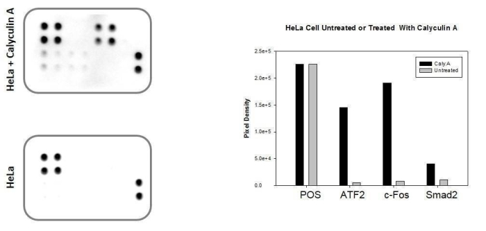
Typical Data
Figure 1 - HeLa cells were grown to 80% confluency and then serum starved overnight. Cells were either untreated (bottom panel) or treated (top panel) with Calyculin A for 30 minutes. Data shown are from a 20 second exposure using a chemiluminescence imaging system.

Storage/Stability
For best results, store the entire kit frozen at -20°C upon arrival. Stored frozen, the kit will be stable for at least 6 months which is the duration of the product warranty period. Once thawed, store array membranes and 1X Blocking Buffer at -20°C and all other reagents undiluted at 4°C for no more than 3 months.
Write Your Own Review