C-Series Human EGFR Phosphorylation Antibody Array 1 Kit. Detects 16 site specific and pan Human EGFRs. Suitable for all liquid sample types but intended for use with cell and tissue lysates.
Product Description
Specifications
| Size | 2 Sample Kit, 4 Sample Kit, 8 Sample Kit |
|---|---|
| Species | Human |
| Quantitative/Semi-Quantitative | Semi-Quantitative |
| Number of Targets Detected | 16 |
| Compatible Sample Types | Cell Culture Supernatants, Plasma, Serum, Tissue Lysates, Cell Lysates |
| Solid Support | Membrane |
| Method Of Detection | Chemiluminescence |
| Design Principle | Sandwich-based |
| Research Area | Post-Translational Modifications, Phosphorylation, HER/ErbB Signaling |
| Estimated Lead Time | 1-2 business days |
| Shipping Type | Blue ice |
| Storage | -20°C |
Risk-Free Guarantee
We offer a 100% guarantee on all ELISA kits and membrane cytokine arrays.
Learn More
Amazon Gift Cards!
$5 Amazon gift card in every kit box purchased.
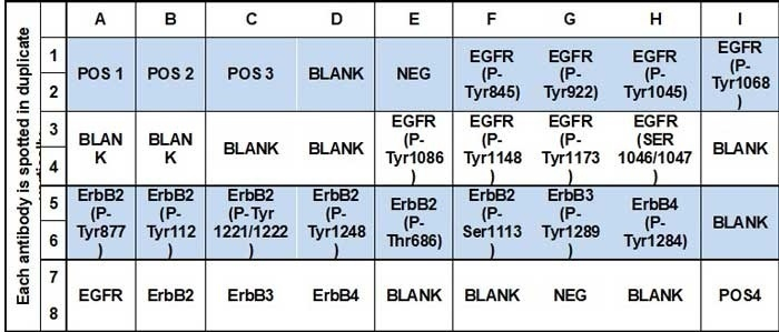
| Scroll over each target protein for more information | ||||
|---|---|---|---|---|
|
EGFR
(P-Tyr845)
|
EGFR
(P-Tyr992)
|
EGFR
(P-Tyr1045)
|
EGFR
(P-Tyr1068)
|
EGFR
(P-Tyr1086)
|
|
EGFR
(P-Tyr1148)
|
EGFR
(P-Tyr1173)
|
EGFR
(P-Ser1046/1047)
|
ErbB2
(P-Tyr877)
|
ErbB2
(P-Tyr1112)
|
|
ErbB2
(P-Tyr 1221/1222)
|
ErbB2
(P-Tyr1248)
|
ErbB2
(P-Thr686)
|
ErbB2
(P-Ser1113)
|
ErbB3
(P-Tyr1289)
|
|
ErbB4
(P-Tyr1284)
|
||||
Application Notes
Suggested Application
Multiplexed Protein Detection; Detection of Relative Protein Expression; Detecting Patterns of Cytokine Expression; Biomarker/ Key Factor Screening; Identifying Key Factors; Confirming a Biological Process
Kit Components
- Human EGFR Phosphorylation Antibody Array C1 Membranes
- Blocking Buffer
- Wash Buffer 1
- Wash Buffer 2
- Biotinylated Detection Antibody Cocktail
- Streptavidin-Conjugated HRP
- Detection Buffer C
- Detection Buffer D
- Lysis Buffer
- Protease Inhibitor Cocktail
- Phosphatase Inhibitor Cocktail Set I
- Phosphatase Inhibitor Cocktail II
- 8-Well Incubation Tray
- Plastic Sheets
- Array Templates
- Manual
Other Materials Required
- Pipettors, pipet tips and other common lab consumables
- Orbital shaker or oscillating rocker
- Tissue Paper, blotting paper or chromatography paper
- Adhesive tape or Saran Wrap
- Distilled or de-ionized water
- A chemiluminescent blot documentation system (such as UVP's ChemiDoc-It® or EpiChem II Benchtop Darkroom or GE's ImageQuant™ LAS 4000 or Amersham Imagers 600 and 680), X-ray Film and a suitable film processor, or another chemiluminescent detection system.
Protocol Outline
- Block membranes
- Incubate with Sample
- Incubate with Biotinylated Detection Antibody Cocktail
- Incubate with HRP-Conjugated Streptavidin
- Incubate with Detection Buffers
- Image with chemiluminescent imaging system
- Perform densitometry and analysis
Storage/Stability
For best results, store the entire kit frozen at -20°C upon arrival. Stored frozen, the kit will be stable for at least 6 months which is the duration of the product warranty period. Once thawed, store array membranes and 1X Blocking Buffer at -20°C and all other reagents undiluted at 4°C for no more than 3 months.
Park JE., Hypoxic Tumor Cell Modulates Its Microenvironment to Enhance Angiogenic and Metastatic Potential by Secretion of Proteins and Exosomes. Molecular & Cellular Proteomics, June 1, 9, 1085-1099.
Species:
Human
Sample Type:
Cell Lysate
Kesanakurti D., et al. Functional cooperativity by direct interaction between PAK4 and MMP-2 in the regulation of anoikis resistance, migration and invasion in glioma. Cell Death and Disease (2012) 3, e445; doi:10.1038/cddis.2012.182
Species:
Human
Sample Type:
Cell Lysate (Glioma Tumor lysate)
Kesanakurti D., Chetty C., Rajasekhar Maddirela D., et al. Functional cooperativity by direct interaction between PAK4 and MMP-2 in the regulation of anoikis resistance, migration and invasion in glioma. Cell Death Dis. 2012 Dec 20;3:e445. doi: 10.1038/cddis.2012.182.
Species:
Human
Sample Type:
Cell Lysate (Human glioma cell lines with siRNA sequences against PAK4)
Cheng KW, Wong CC, Alston N, et al. Aerosol administration of phospho-sulindac inhibits lung tumorigenesis. Mol. Cancer Ther. 2013 May 3; 12: 1417-1428
Species:
Human
Sample Type:
Cell Lysate (A549 cells)
Rios-Doria J., Sabol D., Chesebrough J., et al. A Monoclonal Antibody to ADAM17 Inhibits Tumor Growth by Inhibiting EGFR and Non-EGFR-Mediated Pathways. Mol Cancer Ther. 2015 Jul;14(7):1637-49. doi: 10.1158/1535-7163.MCT-14-1040.
Species:
Human
Sample Type:
Tissue Lysate (Human tumor cells xenograft models)
Mohan N., Shen Y., Endo Y., et al. Trastuzumab, but Not Pertuzumab, Dysregulates HER2 Signaling to Mediate Inhibition of Autophagy and Increase in Reactive Oxygen Species Production in Human Cardiomyocytes. Mol Cancer Ther. 2016 Jun;15(6):1321-31. doi: 10.1158/1535-7163.MCT-15-0741.
Species:
Human
Sample Type:
Cell Lysate (Human Primary Cardiomyocytes)
Zhang J., et al. YAP-dependent induction of amphiregulin identifies a non-cell-autonomous component of the Hippo pathway. Nat Cell Biol. 2009 Dec;11(12):1444-50. doi: 10.1038/ncb1993.
Species:
Human
Sample Type:
Cell Lysate (MCF10A cell lines transfected with agent)
Zhu S., et al. DARPP-32 Increases Interactions Between Epidermal Growth Factor Receptor and ERBB3 to Promote Tumor Resistance to Gefitinib. Gastroenterology. 2011 Nov;141(5):1738-48.e1-2. doi: 10.1053/j.gastro.2011.06.070.
Species:
Human
Sample Type:
Cell Lysate (MKN-28 gastric cancer cell lines)
Giagulli C, Caccuri F, Zorzan S, Bugatti A, Zani A, Filippini F, Manocha E, D'Ursi P, Orro A, Dolcetti R, Caruso A. B-cell clonogenic activity of HIV-1 p17 variants is driven by PAR1-mediated EGF transactivation. Cancer Gene Ther. 2020 Oct 22. doi: 10.1038/s41417-020-00246-9. Epub ahead of print. PMID: 33093643.
Species:
Human
Sample Type:
Cell Lysate
Bhuvaneswari R., et al. Targeting EGFR with photodynamic therapy in combination with Erbitux enhances in vivo bladder tumor response., Mol Cancer. 2009 Nov 2;8:94. doi: 10.1186/1476-4598-8-94.
Species:
Human
Sample Type:
Cell Lysate (MGH bladder cancer cells)
-
Great productErbB Array was used to test expression in whole cell lysates under differential drug conditions. Nice to have many phospho-site test spots for EGFFR and HER2, but only one each for HER3 and HER4. Many control (positive and negative) spots were helpful as well. Would be easier for data analysis if serial dilution spots (top, left three) were designated more specifically on protocol. Overall very helpful product!
from MGH,
on
Great productI used this array to test the cell lysates from epidermoid carcinoma cell line exposed to EGF, it works well.on
Write Your Own Review