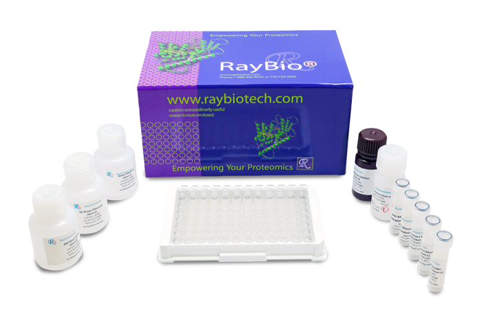
RayBio® Mouse HGF ELISA Kit for cell culture supernatants, plasma, serum, and lysate samples.
Lead time: Typically ships within 1-2 business days. No Friday shipments.
Product Description
Specifications
| Size | 1 Plate Kit, 2 Plate Kit, 5 Plate Kit |
|---|---|
| Species | Mouse |
| Accession Number | Q08048 |
| Gene Id | 15234 |
| Gene Symbols | HGF |
| Protein Name / Synonyms | Hepatocyte growth factor (Hepatopoietin-A) (Scatter factor) (SF) [Cleaved into: Hepatocyte growth factor alpha chain Hepatocyte growth factor beta chain] |
| Quantitative/Semi-Quantitative | Quantitative |
| Specificity | This ELISA kit shows no cross-reactivity with the following cytokines tested: Mouse CD30, L CD30, T CD40, CRG-2, CTACK, CXCL16, Eotaxin , Eotaxin-2, Fas Ligand, Fractalkine, GCSF, GM-CFS, IFN- gamma, IGFBP-3, IGFBP-5, IGFBP-6, IL-1 alpha, IL-1 beta, IL-2, IL-3, IL-3 Rb, IL-4, IL-5, IL-9, IL-10, IL-12 p40/p70, IL-12 p70, IL-13, IL-17, KC, Leptin R, LEPTIN(OB), LIX, L-Selectin, Lymphotactin, MCP-1, MCP- 5, M-CSF, MIG, MIP-1 alpha, MIP-1 gamma, MIP-2, MIP-3 beta, MIP-3 alpha, PF-4, PSelectin, RANTES, SCF, SDF-1 alpha, TARC, TCA-3, TECK, TIMP-1, TNF-alpha, TNF RI, TNF RII, TPO, VCAM-1, VEGF. |
| Compatible Sample Types | Cell Culture Supernatants, Plasma, Serum, Tissue Lysates, Cell Lysates |
| Solid Support | 96-well Microplate |
| Method Of Detection | Colorimetric |
| Design Principle | Sandwich-based |
| Sensitivity | 400 pg/ml Need more sensitivity? Check out the new BIQ-ELISA™ kit for this target. Still not enough? Then your answer is our Ultrasensitive Biomarker Testing Service powered by Simoa™ technology. |
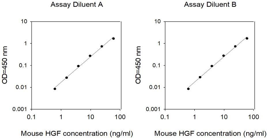
| Detection Range | 400 pg/ml - 60 ng/ml |
| Recommended Dilution (Serum/Plasma) | 2 - 10 fold |
| Estimated Lead Time | 1-2 business days |
| Shipping Type | Blue ice |
| Storage | ≤-20°C |
Risk-Free Guarantee
We offer a 100% guarantee on all ELISA kits and membrane cytokine arrays.
Learn More
Amazon Gift Cards!
$5 Amazon gift card in every kit box purchased.
Hamdi, Hadhami, et al. "Efficacy of epicardially delivered adipose stroma cell sheets in dilated cardiomyopathy." Cardiovascular research 99.4 (2013): 640-647.
Species:
Mouse
Sample Type:
Tissue Lysate (dilated cardiomyopathy model)
Juffer P., Jaspers RT., Lips P., et al. Expression of muscle anabolic and metabolic factors in mechanically loaded MLO-Y4 osteocytes. Am J Physiol Endocrinol Metab. 2012 Feb 15;302(4):E389-95. doi: 10.1152/ajpendo.00320.2011.
Species:
Mouse
Sample Type:
Conditioned Media (MLO-Y4 osteocytes cells)
Hora, Caroline, Pamela Romanque, and Jean?François F. Dufour. "Effect of sorafenib on murine liver regeneration." Hepatology 53.2 (2011): 577-586.
Species:
Mouse
Sample Type:
Tissue lysate
Rehm, A. et al. Dendritic cell-mediated survival signals inEm-Myc B-cell lymphoma depend on the transcription factor C/EBPb. Nat. Commun.5:5057 doi: 10.1038/ncomms6057 (2014).
Species:
Human
Sample Type:
Conditioned Media
Li L., Black R., Ma Z., et al. Use of mouse hematopoietic stem and progenitor cells to treat acute kidney injury. Am J Physiol Renal Physiol. 2012 Jan 1;302(1):F9-F19. doi: 10.1152/ajprenal.00377.2011.
Species:
Mouse
Sample Type:
Conditioned Media
Xinaris, Christodoulos, et al. "A novel strategy to enhance mesenchymal stem cell migration capacity and promote tissue repair in an injury specific fashion." Cell Transplantation 22.3 (2013): 423-436.
Species:
Mouse
Sample Type:
Conditioned Media (MSCs (AKI injury repair model))
Huey K., Smith S., Sulaeman A., Breen E. Skeletal myofiber VEGF is necessary for myogenic and contractile adaptations to functional overload of the plantaris in adult mice. J Appl Physiol (1985). 2016 Jan 15;120(2):188-95. doi: 10.1152/japplphysiol.00638.2015.
Species:
Mouse
Sample Type:
Tissue Lysate (Skeletal muscle lysate from plantaris muscle)
Sato C., Iwasaki T., Kitano S., et al. Sphingosine 1-phosphate receptor activation enhances BMP-2-induced osteoblast differentiation. Biochem Biophys Res Commun. 2012 Jun 22;423(1):200-5. doi: 10.1016/j.bbrc.2012.05.130.
Species:
Mouse
Sample Type:
Conditioned Media (C2C12 cells, mouse myoblast cell line and role of S1P)
DÃaz-Coránguez M, Segovia J, López-Ornelas A, et al. Transmigration of Neural Stem Cells across the Blood Brain Barrier Induced by Glioma Cells. PLoS ONE. 2013;8(4):e60655. doi:10.1371/journal.pone.0060655
Species:
Rat
Sample Type:
Conditioned Media (astrocytes, Glioma C6)
Talbot N., Sparks W., Powell A., et al. Quantitative and semiquantitative immunoassay of growth factors and cytokines in the conditioned medium of STO and CF-1 mouse feeder cells. In Vitro Cell Dev Biol Anim. 2012 Jan;48(1):1-11. doi: 10.1007/s11626-011-9467-7
Species:
Mouse
Sample Type:
Conditioned Media (Primary CF-1 fibtoblasts from 13.5d fetsus of CF-1 mice, and STO fibroblasts)
Write Your Own Review


