Mouse Apoptosis Array C1
Membrane-based antibody array for detection of 38 mouse apoptosis biomarkers, including Caspase 3, Caspase 8, bad, bax, bcl-2, etc.
Product Description
Specifications
| Size | 2 Sample Kit, 4 Sample Kit, 8 Sample Kit |
|---|---|
| Species | Mouse |
| Quantitative/Semi-Quantitative | Semi-Quantitative |
| Number of Targets Detected | 38 |
| Compatible Sample Types | Cell Culture Supernatants, Plasma, Serum, Tissue Lysates, Cell Lysates |
| Solid Support | Membrane |
| Method Of Detection | Chemiluminescence |
| Design Principle | Sandwich-based |
| Research Area | Apoptosis, Akt Signaling, IGF Signaling, MAPK Signaling, PI3K-AKT Signaling, p53 Signaling, NFkB Signaling |
| Estimated Lead Time | 1-2 business days |
| Shipping Type | Blue ice |
| Storage | -20°C |
Risk-Free Guarantee
We offer a 100% guarantee on all ELISA kits and membrane cytokine arrays.
Learn More
Amazon Gift Cards!
$5 Amazon gift card in every kit box purchased.
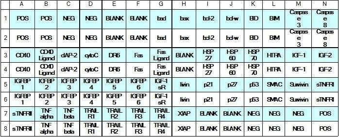
| Scroll over each target protein for more information | ||||
|---|---|---|---|---|
|
bad
|
bax
|
bcl-2
|
bcl-w
|
BID
|
|
BIM
|
Caspase-3
|
Caspase-8
|
CD40
(TNFRSF5)
|
CD40 Ligand
(TNFSF5)
|
|
cIAP-2
|
Cytochrome C
|
DR6
(TNFRSF21)
|
Fas
(TNFRSF6/Apo-1)
|
Fas Ligand
(TNFSF6)
|
|
HSP27
|
HSP60
|
HSP70
|
HTRA2
|
IGFBP-1
|
|
IGFBP-2
|
IGFBP-3
|
IGFBP-4
|
IGFBP-5
|
IGFBP-6
|
|
IGF-1
|
IGF-2
|
p27/Kip1
(CDKN1B)
|
p53
|
|
|
SMAC
|
Survivin
(BIRC5)
|
TNF RI
(TNFRSF1A)
|
TNF RII
(TNFRSF1B)
|
TNF alpha
|
|
TNF beta
(TNFSF1B)
|
TRAIL R2
(TNFRSF10B/DR5)
|
XIAP
|
||
Application Notes
Suggested Application
Multiplexed Protein Detection; Detection of Relative Protein Expression; Detecting Patterns of Cytokine Expression; Biomarker/ Key Factor Screening; Identifying Key Factors; Confirming a Biological Process
Kit Components
- Mouse Apoptosis Antibody Array C1 Membranes
- Blocking Buffer
- Wash Buffer 1
- Wash Buffer 2
- Biotinylated Detection Antibody Cocktail
- Streptavidin-Conjugated HRP
- Detection Buffer C
- Detection Buffer D
- Lysis Buffer
- Protease Inhibitor Cocktail
- 8-Well Incubation Tray
- Plastic Sheets
- Array Templates
- Manual
Other Materials Required
- Pipettors, pipet tips and other common lab consumables
- Orbital shaker or oscillating rocker
- Tissue Paper, blotting paper or chromatography paper
- Adhesive tape or Saran Wrap
- Distilled or de-ionized water
- A chemiluminescent blot documentation system (such as UVP's ChemiDoc-It® or EpiChem II Benchtop Darkroom or GE's ImageQuant™ LAS 4000 or Amersham Imagers 600 and 680), X-ray Film and a suitable film processor, or another chemiluminescent detection system.
Protocol Outline
- Block membranes
- Incubate with Sample
- Incubate with Biotinylated Detection Antibody Cocktail
- Incubate with HRP-Conjugated Streptavidin
- Incubate with Detection Buffers
- Image with chemiluminescent imaging system
- Perform densitometry and analysis
Storage/Stability
For best results, store the entire kit frozen at -20°C upon arrival. Stored frozen, the kit will be stable for at least 6 months which is the duration of the product warranty period. Once thawed, store array membranes at -20°C and all other reagents undiluted at 4°C for no more than 3 months.
-
Good and easy methodThe method is easy and simple. The detection method is compatible with other detection system. I used IRDye 800CW Streptavidin instead of HRP-Streptavidin provided in the kit and scanned the membranes with LI-COR Odyssey system. The method works and results shows pretty good.
from University of Missouri-Kansas city,
on
Great productThe kit worked but the spot for TNF alpha did not show up at all in both my wildtype and the experimental condition. Considering the fact that I used 12 month old mouse brain tissue and TNF alpha is expressed there.So, I was able to probe only 29/30 antibodies.from Northwestern University,
on
Write Your Own Review