Introduction
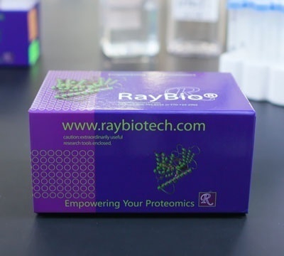
Protein phosphorylation plays an unusually prominent role in cell signaling, development and growth. The RayBio® G-Series Human Pathway Explorer Phosphorylation Antibody Array 1 is a very rapid, convenient, and sensitive assay that can simultaneously detect multiple protein phosphorylations and be used to monitor the activation or function of important biological pathways.
RayBiotech is committed to develop a series of phosphorylation antibody arrays. RayBio® Human Pathway Explorer Phosphorylation Antibody Array 1 is specifically designed for simultaneous identification of the relative levels of phosphorylation of 393 different human signaling pathway proteins in cell lysates. By monitoring the changes in protein phosphorylation in your experimental model system, you can verify pathway activation in your cell lines without spending excess time and effort performing an analysis of immunoprecipitation and/or Western Blot.
To use the RayBio® G-Series Human Pathway Explorer Phosphorylation Antibody Array 1, treated or untreated cell lysate is added into antibody array glass slide wells. The antibody array slide wells are washed, and biotinylated anti-phospho Tyr/Thr/Ser antibodies are then used to detect the phosphorylated Tyrosine/Threonine/Serine on target proteins. After incubation with a fluorescent dye-conjugated streptavidin (Cy3 equivalent), the slides can then be imaged using a laser scanner, such as the Axon GenePix, using the Cy3 channel.
Product Features
- High sensitivity and specificity
- Low sample volume (10-100 µl per array)
- Large dynamic range of detection
- Compatible with most sample types
- Test 4 or 8 samples on each slide
- Suitable for high-throughput assays
Target Names
14-3-3 zeta, 14-3-3b, 4E-BP1, AB 1-40, ABL1, ACETYL CoA, ACK1, ADD1, AKT, AKT1S1, ALK, AMPK a1, AMPK b1, AR, ARAF, ARHGEF2, ASK1, ATF1, ATF2, ATF4, ATM, ATR, ATRIP, AURKA, AURKB, AURKC, AXL, BAD, BAX, BCL-2, BCL-XL, BCR, Beclin1, beta-Arrestin 1, beta-Catenin, BID, BIM, BLK, BLNK, BMX, B-raf, BRCA1, BTK, CAMK1, CAMK2A, CAMK2d, CAMK4, Casein Kinase 1a, Casein Kinase 2b, Caspase 1, Caspase 2, Caspase 3, Caspase 6, Caspase 7, Caspase 8, Caspase 9, Catalase, Caveolin-1, CBL, CD19, CD247, CD28, CD32, CD4, CD45, CD5, CD61, CDC25A, CDC25B, CDC25C, CDC37, CDK1, CDK2, CDK5, CDK7, c-Fos, CHK1, CHK2, c-Jun, Claudin-3, CLDN6, CLDN7, c-Myc, Cofilin, Cortactin, CPI17 alpha, CREB1, c-Rel, CRK, CRKL, CTNND1, CXCR4, Cyclin B1, Cyclin D1, Cyclin D2, Cyclin E1, Cytokeratin 18, Cytokeratin 8, DAB1, DAPP1, DAXX, DDR1, DDX5, DNM1, DOK1, DTK, DUSP1, E2F1, EEF-2, EGFR, EIF2a, EIF4B, EIF4E, EIF4G1, ELK1, eNOS, EPB41, EphA1, EphA2, EphA3, EPHA4, EphA5, EphA6, EphA7, EphA8, EphB1, EphB2, EphB3, EphB4, EphB6, Ephrin B2, EPO R, ERa, ErbB2, ErbB3, ErbB4, ERK1/2, ERK2, Ezrin, Factor 3, FADD, FAK, FAS, FER, FGFR1, FGFR2, FGFR2 (a isoform), FGR, Filamin A, Flt-3, FosB, FoxO1, Foxo3, FRK, FRS2, FYN, G3BP, GAB2, GABRB1, GAP43, GATA-1, GJA1, GRIA1, GRIA2, GRIN1, GRIN2A, Grin2b, GRK1, GRK2, GSK3a, GSk3b, HCK, HDAC1, HDAC2, HDAC3, HDAC4, HDAC5, HDAC6, HDAC8, His H2A.x, Histone H3, HNF-4a, HRS, HSF1, HSL, HSP27, HSP90, ICAM-1, ICK, IFN-a/b R1, IFN-gamma R1, IGF-1 R, IGFBP-3, IGF-II R, IkB beta, IkB epsilon, IkBa, IKK gamma, IKKa, IKKb, IL-10 R alpha, IL-13 Ra1, IL-2 sRa, IL-3 R alpha, IL-4 R, IL-7 R alpha, iNOS, Insulin R, IntegrinB1, IRF3, IRS1, ITGB4, ITK, JAK1, JAK2, JAK3, JNK, JunB, JunD, KCNA3, KCNB1, KCNIP3, KSR1, LaminA+C, LAT, LCK, LIMK1, LKB1, LSD1, LTK, Lyn, MAP3K1, MAP3K8, MAPK6, MARCKS, MATK, M-CSF R, MDM2, MDM4, MEF2A, MEF2C, MEF2D, MEK1, MEK2, MET, MITF, MKK3, MKK4, MKK6, MKK7, MKNK1/MNK1, MLKL, MSK1, MSK2, MSP/MST1, mTOR, MUCI, MUSK, MYPT1, Nbs1, NF2, NFATC3, NFATC4, NF-KB (P65), NFkb2, NGFR, NR3C3, P130Cas, P21, P27, P300, P38, P53, P63, P70S6K, PAK1, PAK2, PAK3, PAK4, PARP, Paxillin, PDGF Ra, PDGF Rb, PDK1, PEA15, PECAM-1, PIM1, PIP5K3, PKC a, PKC d, PKC epsilon, PKC theta, PKC zeta, PKD1, PKD2, PKMYT1, PKR, PLA2G4A, PLCB3, PLCg1, PLCG2, PLD1, PLD2, PLK1, PP2A, PPARg2, PPP1R1B, PRKACA, PRKCB, PTEN, PTK6, PYK2, Rac1, RACGAP1, RAD51, Raf1, RASGRF1, RB1, RELB, Ret, RGS16, RhoA, Rictor, ROR1, ROR2, ROS, RPS6, RPS6KA3, RSK1, RYK, RYR2, SCF R, SHC, SHIP1, SHP-1, SHP-2, Siglec-2, SLP-76/LCP2, Smad1, Smad2, Smad2/3, smad4, Smad5, SMC1, SNCA, SP1, SRC, SREBF1, SRMS, STAT1, STAT2, STAT3, STAT4, STAT5, STAT5b, Stat6, STMN1, Survivin, SYK, SYN1, SYT1, TAK1, Tau, Tec, TGF-B2, TGF-beta RI, Tie-1, Tie-2, TLK1, TNK1, TOP2A, TrkA, TrkB, Tuberin / TSC2, TXK, TYH, TYK2, TYRO10 (DDR2), ULK1, VASP, VAV1, VAV2, VE-Cadherin, VEGF-R1, VEGF-R2, VEGFR3, Vinculin, WASF1, WASP, WNK1, WWOX, XIAP, ZAP70
Application Notes
Suggested Application
Multiplexed Protein Detection; Detection of Relative Protein Expression; Detecting Patterns of Cytokine Expression; Biomarker/ Key Factor Screening; Identifying Key Factors; Confirming a Biological Process
Kit Components
- Human Phosphorylation Antibody Array Slide(s)
- Blocking Buffer
- Wash Buffer 1
- Wash Buffer 2
- Biotinylated Detection Antibody Cocktail
- Streptavidin-Conjugated Fluor
- Lysis Buffer
- Protease Inhibitor Cocktail
- Phosphatase Inhibitor Cocktail II
- Adhesive Plastic Strips
- 30 ml Centrifuge Tube
- Manual
Other Materials Required
- Small plastic boxes or containers
- Pipettors, pipette tips and other common lab consumables
- Orbital shaker or oscillating rocker
- Aluminum foil
- Gene microarray scanner or similar laser fluorescence scanner
Protocol Outline
- Dry the glass slide
- Block array surface
- Incubate with Sample
- Incubate with Biotinylated Detection Antibody Cocktail
- Incubate with Streptavidin-Conjugated Fluor
- Disassemble the glass slide
- Scan with a gene microarray laser scanner
- Perform densitometry and analysis


