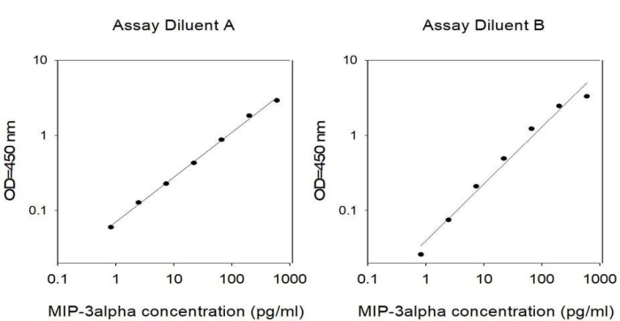
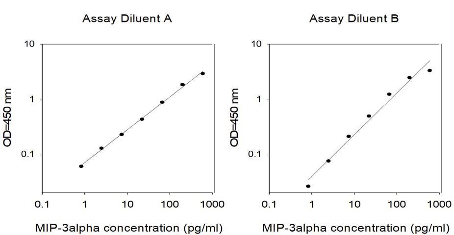
RayBio® Human MIP-3 alpha (CCL20) ELISA Kit for cell culture supernatants, plasma, serum, and lysate samples.
Lead time: Typically ships within 1-2 business days. No Friday shipments.
Product Description
Specifications
| Size | 1 Plate Kit, 2 Plate Kit, 5 Plate Kit |
|---|---|
| Species | Human |
| Accession Number | P78556 |
| Gene Id | 6364 |
| Gene Symbols | CCL20|MIP3A|SCYA20|LARC |
| Protein Name / Synonyms | C-C motif chemokine 20 (Beta-chemokine exodus-1) (CC chemokine LARC) (Liver and activation-regulated chemokine) (Macrophage inflammatory protein 3 alpha) (MIP-3-alpha) (Small-inducible cytokine A20) [Cleaved into: CCL20(1-67) CCL20(1-64) CCL20(2-70)] |
| Quantitative/Semi-Quantitative | Quantitative |
| Specificity | This ELISA kit shows no cross-reactivity with any of the cytokines tested: Human Angiogenin, BDNF, BLC, ENA-78, FGF-4, IL-1 alpha, IL-1 beta, IL-2, IL-3, IL-4, IL-5, IL-7, IL-8, IL-9, IL-10, IL-11, IL-12 p70, IL-12 p40, IL-13, IL-15, I-309, IP-10, G-CSF, GM-CSF, IFN-gamma, Leptin, MCP-1, MCP-2, MCP-3, MDC, MIP-1 alpha, MIP-1 beta, MIP-1 delta, PARC, PDGF, RANTES, SCF, TARC, TGF-beta, TIMP-1, TIMP-2, TNF-alpha, TNF-beta, TPO, VEGF. |
| Compatible Sample Types | Cell Culture Supernatants, Plasma, Serum, Tissue Lysates, Cell Lysates |
| Solid Support | 96-well Microplate |
| Method Of Detection | Colorimetric |
| Design Principle | Sandwich-based |
| Sensitivity | 1.5 pg/ml Need more sensitivity? Check out the new BIQ-ELISA™ kit for this target. Still not enough? Then your answer is our Ultrasensitive Biomarker Testing Service powered by Simoa™ technology. |
| Detection Range | 1.5 pg/ml - 600 pg/ml |
| Recommended Dilution (Serum/Plasma) | 2 fold |
| Estimated Lead Time | 1-2 business days |
| Shipping Type | Blue ice |
| Storage | ≤-20°C |
Risk-Free Guarantee
We offer a 100% guarantee on all ELISA kits and membrane cytokine arrays.
Learn More
Amazon Gift Cards!
$5 Amazon gift card in every kit box purchased.
Wang J., Su X., Yang L., et al. The influence of myeloid-derived suppressor cells on angiogenesis and tumor growth after cancer surgery. Int J Cancer. 2016 Jun 1;138(11):2688-99. doi: 10.1002/ijc.29998.
Species:
Human
Sample Type:
Serum (Lung Cancer Patients)
Crean D., Cummins E., Bahar B., et al. Adenosine Modulates NR4A Orphan Nuclear Receptors To Attenuate Hyperinflammatory Responses in Monocytic Cells. J Immunol. 2015 Aug 15;195(4):1436-48. doi: 10.4049/jimmunol.1402039.
Species:
Human
Sample Type:
Conditioned Media (THP-1 macrophage line)
Leea Y,Chioub T, Tzengd W, Chua S. Macrophage inflammatory protein-3 influences growth of K562 leukemia cells in co-culture with anticancer drug-pretreated HS-5 stromal cells. Toxicology. 2008;249:116-122.
Species:
Human
Sample Type:
Conditioned Media (conditioned medium)
Leea Y,Chioub T,Tzengd W,Chua S.Macrophage inflammatory protein-3 influences growth of K562 leukemiacells in co-culture with anticancer drug-pretreated HS-5 stromal cells.Toxicology.2008;249:116-122
Species:
Human
Sample Type:
Conditioned Media
Iwata T, Tanaka K, Inoue Y, et al. Macrophage inflammatory protein-3 alpha (MIP-3a) is a novel serum prognostic marker in patients with colorectal cancer. J Surg Oncol. 2013 Feb;107(2):160-166.
Species:
Human
Sample Type:
Serum
Involvement of corneal epithelial cells in the Th17 response in an in vitro bacterial inflammation model
Species:
Human
Sample Type:
Conditioned Media (corneal epithelial cell (eye))
Write Your Own Review


