Product Description
Specifications
| Size | 2 Sample Kit, 4 Sample Kit, 8 Sample Kit |
|---|---|
| Species | Human |
| Quantitative/Semi-Quantitative | Semi-Quantitative |
| Number of Targets Detected | 19 |
| Compatible Sample Types | Cell Culture Supernatants, Plasma, Serum, Tissue Lysates, Cell Lysates |
| Solid Support | Membrane |
| Method Of Detection | Chemiluminescence |
| Design Principle | Sandwich-based |
| Research Area | Apoptosis, Akt Signaling, HIF-1 alpha Signaling, IGF Signaling, JAK/STAT Signaling, MAPK Signaling, mTOR Signaling, PI3K-AKT Signaling, TGF-beta Signaling, Wnt/beta-Catenin Signaling, p53 Signaling, NFkB Signaling |
| Estimated Lead Time | 1-2 business days |
| Shipping Type | Blue ice |
| Storage | -20°C |
Risk-Free Guarantee
We offer a 100% guarantee on all ELISA kits and membrane cytokine arrays.
Learn More
Amazon Gift Cards!
$5 Amazon gift card in every kit box purchased.
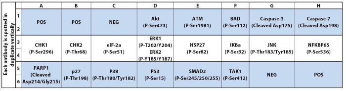
| Scroll over each target protein for more information | ||||
|---|---|---|---|---|
|
Akt (P-Ser473)
(PKB (P-Ser473))
|
ATM (P-Ser1981)
|
BAD (P-Ser112)
|
Caspase-3 (Cleaved Asp175)
|
Caspase-7 (Cleaved Asp198)
|
|
CHK1 (P-Ser296)
|
CHK2 (P-Thr68)
|
eIF-2a (P-Ser52)
|
ERK1 (P-T202/Y204)/ERK2 (P-Y185/Y187)
|
HSP27 (P-Ser82)
|
|
IKBa (P-Ser32)
|
JNK (P-Thr183/Tyr185)
|
NFKBP65 (P-Ser536)
|
PARP1 (Cleaved Asp214/Gly215)
|
p27 (P-Thr198)
|
|
p38 (P-Thr180/Tyr182)
|
P53 (P-Ser15)
|
SMAD2 (Ser245/250/255)
|
TAK1 (P-Ser412)
|
|
Typical Results


Application Notes
- Human Apoptosis Signaling Antibody Array C1 Membranes
- Blocking Buffer
- Detection Antibody Cocktail
- 1,000X HRP-Anti-Rabbit IgG Concentrate
- 20X Wash Buffer I Concentrate
- 20X Wash Buffer II Concentrate
- 2X Cell Lysis Buffer Concentrate
- Detection Buffer C
- Detection Buffer D
- 8-Well Incubation Tray w/ Lid
- Protease Inhibitor Cocktail
- 100x Phosphatase Inhibitor Cocktail I
- Phosphatase Inhibitor Cocktail II
- Plastic Sheets
- Array Map Template
- Manual
- Pipettors, pipet tips and other common lab consumables
- Orbital shaker or oscillating rocker
- Tissue Paper, blotting paper or chromatography paper
- Adhesive tape or Saran Wrap
- Distilled or de-ionized water
- A chemiluminescent blot documentation system (such as UVP's ChemiDoc-It® or EpiChem II Benchtop Darkroom or GE's ImageQuant™ LAS 4000 or Amersham Imagers 600 and 680), X-ray Film and a suitable film processor, or another chemiluminescent detection system.
- Block membranes
- Incubate with Sample
- Incubate with Detection Antibody Cocktail
- Incubate with HRP-Conjugated anti-IgG
- Incubate with Detection Buffers
- Image with chemiluminescent imaging system
- Perform densitometry and analysis
Storage/Stability
Vergani E, Dugo M, Cossa M, Frigerio S, Di Guardo L, Gallino G, Mattavelli I, Vergani B, Lalli L, Tamborini E, Valeri B, Gargiuli C, Shahaj E, Ferrarini M, Ferrero E, Gomez Lira M, Huber V, Vecchio MD, Sensi M, Leone BE, Santinami M, Rivoltini L, Rodolfo M, Vallacchi V. miR-146a-5p impairs melanoma resistance to kinase inhibitors by targeting COX2 and regulating NFkB-mediated inflammatory mediators. Cell Commun Signal. 2020 Sep 23;18(1):156. doi: 10.1186/s12964-020-00601-1. PMID: 32967672; PMCID: PMC7510138.
Hii, LW., Lim, SH.E., Leong, CO. et al. The synergism of Clinacanthus nutans Lindau extracts with gemcitabine: downregulation of anti-apoptotic markers in squamous pancreatic ductal adenocarcinoma. BMC Complement Altern Med 19, 257 (2019). https://doi.org/10.1186/s12906-019-2663-9
Smolensky D, Rhodes D, McVey DS, et al. High-Polyphenol Sorghum Bran Extract Inhibits Cancer Cell Growth Through ROS Induction, Cell Cycle Arrest, and Apoptosis. J Med Food. 2018;21(10):990-998. doi:10.1089/jmf.2018.0008
Bao X, Shi J, Xie F, et al. Proteolytic Release of the p75NTR Intracellular Domain by ADAM10 Promotes Metastasis and Resistance to Anoikis. Cancer Res. 2018;78(9):2262-2276. doi:10.1158/0008-5472.CAN-17-2789
-
Great productWas investigating ulcerative colitis serum exomes and drug ADS04 on HPECs and any downstream apoptosis. The kit was easy to use and gave a really good, clean output at the end
from UCLA,
on
Great productThe human apoptosis array provides a fast and comprehensive glance about the key signaling involved in the apoptotic pathway. It's a one shot experiment with certain controls already on the blot, pretty simple to execute and normalize. Would recommend it.from Moffitt Cancer Center,
on
Great productThis apoptosis kit was very user-friendly and gave detailed instructions on how to analyze findings using a software like Excel. This assay was very helpful for examining multiple gene targets simultaneously, so we got a lot of data out of 1 experiment. Negative and positive controls were included on the blots, which was useful in subsequent analyses.Notes: the Caspase3 antibody seemed a lot more sensitive than other antibodies on the blot. Also, some of the gene names in the kit manual differed from those listed on the website, so keep that in mind when analyzing data to ensure that the correct gene is being referred to.from Medical College of Wisconsin,
on
Great productIt seems antibodies are not evenly attached on the membrane. Some signals show up outside of spots. It has high background when I follow up the suggested procedures. I cannot see any benefit of this array compared to other array such as apoptosis array or other company's similar product.from Penn State College of Medicine,
on
Write Your Own Review