ELISA Kits
For all your sensitive and quantitative detection needs - whether you're measuring low antigen concentrations, analyzing cellular regulation, conducting quality control studies, or tackling another challenge - RayBiotech is here to support you. With ELISA Kits that blend high-quality performance with competitive pricing and next-day shipping, you’ll generate more reliable data for confident decision-making.

Choose RayBiotech ELISA Kits for Affordable Performance
Helping scientists generate data quickly and confidently is at the heart of what we do at RayBiotech, and one reason we’ve built such a large number of targets and validated antibody pairs. With over 6,000 targets from 17 different species, you’re bound to find a ready-made reagent to get your studies off the ground quickly.
But a reagent is only as good as the care that goes into its manufacturing and quality control, which is why we manufacture all ELISA Kits on-site in our facility in Peachtree Corners, Georgia, USA. From the production and validation of antibody pairs that recognize a wide variety of soluble proteins (cytokines, growth factors, signaling molecules), transcription factors, and post-translational modifications to the buffers and related reagents needed to complete the ELISA, we conduct rigorous quality control and validation studies so you can be confident in our data.
ELISA Kit Categories
Popular ELISA Kits
-
 Human IL-6 ELISA Kit
The Human IL-6 ELISA is a pre-coated sandwich-based assay kit for the measurement of Interleukin 6 (IL6) in human cell culture supernatants, plasma, serum, and lysate samples.
Lead time: Typically ships within 1-2 business days. No Friday shipments.
-
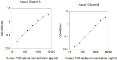 Human TNF alpha ELISA Kit RayBio® Human TNF alpha ELISA Kit for cell culture supernatants, plasma, serum, and lysate samples. 30 pg/ml sensitivity.
 -
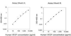 Human VEGF-A ELISA Kit
RayBio® Human VEGF-A ELISA Kit for cell culture supernatants, plasma, serum, and lysate samples.
Lead time: Typically ships within 1-2 business days. No Friday shipments.
-
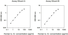 Human IL-1 beta ELISA Kit
RayBio® Human IL-1 beta (IL-1 F2) ELISA Kit for cell culture supernatants, plasma, and serum samples.
Lead time: Typically ships within 1-2 business days. No Friday shipments.
-
 Mouse IL-6 ELISA Kit
RayBio® Mouse IL-6 ELISA Kit for cell culture supernatants, plasma, serum, and lysate samples.
Lead time: Typically ships within 1-2 business days. No Friday shipments.
-
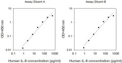 Human IL-8 ELISA Kit
RayBio® Human IL-8 (CXCL8) ELISA Kit for cell culture supernatants, plasma, serum, and lysate samples.
Lead time: Typically ships within 1-2 business days. No Friday shipments.
-
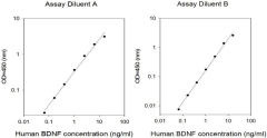 Human BDNF ELISA Kit
The RayBiotech BDNF ELISA kit is an in vitro enzyme-linked immunosorbent assay for the quantitative measurement of human BDNF in serum, plasma, and cell culture supernatant samples.
Lead time: Typically ships within 1-2 business days. No Friday shipments.
New Products
-
 m7G (N7-methylguanosine) ELISA Kit The RayBiotech m7G ELISA Kit is a competitive enzyme immunoassay (EIA) developed for rapid detection and quantitation of m7G in urine, serum, plasma, and RNA samples collected from human, rat or mouse.
 -
 m1A (N1-methyladenosine) ELISA Kit The RayBio® m1A ELISA Kit is a competitive enzyme immunoassay developed for rapid detection and quantitation of m1A in urine, serum, or plasma samples collected from human, rat, or mouse.
 -
 Alpha-MSH ELISA Kit The RayBio® Alpha-MSH Enzyme Immunoassay (EIA) Kit is an in vitro quantitative assay for detecting human, mouse, or rat melanocyte-stimulating hormone alpha based on the competitive ELISA principle. The immunoassy is optimized for serum, plasma and cell culture media.
 -
Phospho-CDC25A (S293) and Total CDC25A ELISA Kit The RayBio® Human and Mouse Phospho-CDC25A (S293) and Total CDC25A ELISA Kit semi-quantitatively measures phosphorylated CDC25A (Ser293) and total CDC25A in lysate samples.
-
Human ATF-1 Activity Assay Kit The RayBio® ATF-1 Transcription Factor Activity Assay Kit uses a dsDNA coated plate with canonical ATF-1 binding sequences to semi-quantitatively detect active ATF-1 in lysates or nuclear extracts. Dry ice shipment (additional fee).
-
Human BTLA / HVEM Binding Assay Kit The Human BTLA / HVEM Binding Assay Kit uses a rapid, simple, and sensitive method to characterize the binding affinity between herpes virus entry mediator and its receptor B- and T-lymphocyte attenuator in the presence of potential inhibitors within 4 hours.
4-8 week development phase.
What customers are saying
“Amazing kit [Mouse Copeptin EIA, Cat# EIAM-COP]. Easy to handle, convenient based on incubation time. Highly sensitive, gives me appropriate results. Awesome product!!!“
Purnima
University of Tennessee Health Science Center
“The Phospho-PTEN (S380)/ total PTEN ELISA [CAT# PEL-PTEN-S380-T] works very well and gives repetitive and accurate results with lysates from cancer cell lines.”
Marina
Australis Pharmaceuticals

Featured Service
ELISA Services
Maximize operational efficiency while receiving data you can depend on when you trust your ELISA service needs to the experienced scientists at RayBiotech.
Rigorous Testing for Quality You Can Trust
At RayBiotech, we take our product quality very seriously. We apply a battery of rigorous product-specific quality control tests to ensure your experiments are set up to produce high-quality results.
Reproducibility and Precision
We evaluate intra-assay reproducibility on sandwich ELISA kits by running 2-3 positive control samples in duplicate on a single plate (maximum tolerance = 10% CV). We use 2-3 positive control samples and a full standard curve (maximum tolerance = 12% CV) in at least 2 independent experiments. Our lot-to-lot consistency is tested by comparing calculated concentrations of the 2-3 positive control samples with calibration curves of the current lot and previous lot (maximum tolerance = 20% CV).
Dilution Linearity and Recovery
We determine recovery by spiking various levels of target protein into biological samples. Our dilution linearity is tested by performing 2-fold and 4-fold dilutions. Recoveries typically range from 80-130% (maximum tolerance 70-150%). The biological samples tested include serum, EDTA plasma, citrate plasma, heparin plasma (normal healthy donors), and cell culture medium (DMEM or 1640). This testing series determines our recommended dilution ranges for serum and plasma. For lysate-specific sandwich ELISAs (catalog numbers ending in “-CL”), sample types include cell lysates and tissue homogenates of various origins.
Four Steps to Antigen Analysis
Each type of ELISA works a little differently, but they all follow the same basic process:

Plate coating
First, antigens are directly or indirectly immobilized on the surface of micropates.
Plate Blocking
Next, a blocking buffer containing an irrelevant protein is added to bind to the remaining surfaces.
Antibody Incubation
Then, an antigen-specific antibody is added. It binds to the antigens during the incubation step.
Detection
Finally, the signal generated by the direct or secondary tag on the antibody is detected and measured.
Frequently Asked Questions
Still have questions?
Still have questions?